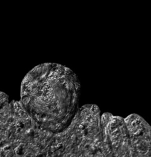
DIC micrograph of a malaria oocyst in the gut of a mosquito (Plasmodium berghei). The malaria parasite is ingested by the mosquito when it feeds on infected blood. The parasite enters the midgut where it undergoes rounds of replication and transformation, before passing through the midgut epithelium. Upon emerging into the insect hemocoel (body cavity), contact with the basal lamina turns the parasite into an oocyst. After the oocyst has matured (a period of weeks) tens of thousands of sporozoites are released into the open circulatory system of the insect. Using a type of motility known as gliding, the sporozoites locate and enter the mosquito salivary gland. They remain here until injected into a mammalian host during the next blood feed, completing the mosquito stage of the parasite life cycle. DIC micrograph; x 40; oocyst is approximately 50 micrometers in length.