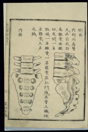
Woodblock illustration from a work on 'Chinese' medicine by the 18th century Japanese physician Hara Masakatsu, published in 1807 (4th year of the Bunkwa era) -- lateral and internal view of the bones of the lumbar and pelvic regions of the skeleton. The hipbone (kegu, also known as kuangu), tailbone (jiangu) and sacrum (kaogu) are shown at the base of the spine. The coccyx or sacrococcyx, shown here, is variously known as weidigu (sacral tail bone), weilü (tail neighbourhood), diduan (end of the sacrum), juegu (protruding bone) and qionggu (poor bone).