Attribution 4.0 International (CC BY 4.0)
You can use this work for any purpose, including commercial uses, without restriction under copyright law. You should also provide attribution to the original work, source and licence. Read more about this licence.
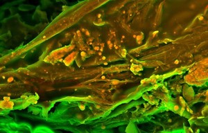
Credit
Human heart (mitral valve) tissue displaying calcification. Sergio Bertazzo, Department of Materials, Imperial College London. Attribution 4.0 International (CC BY 4.0). Source: Wellcome Collection.