Stories
- Article
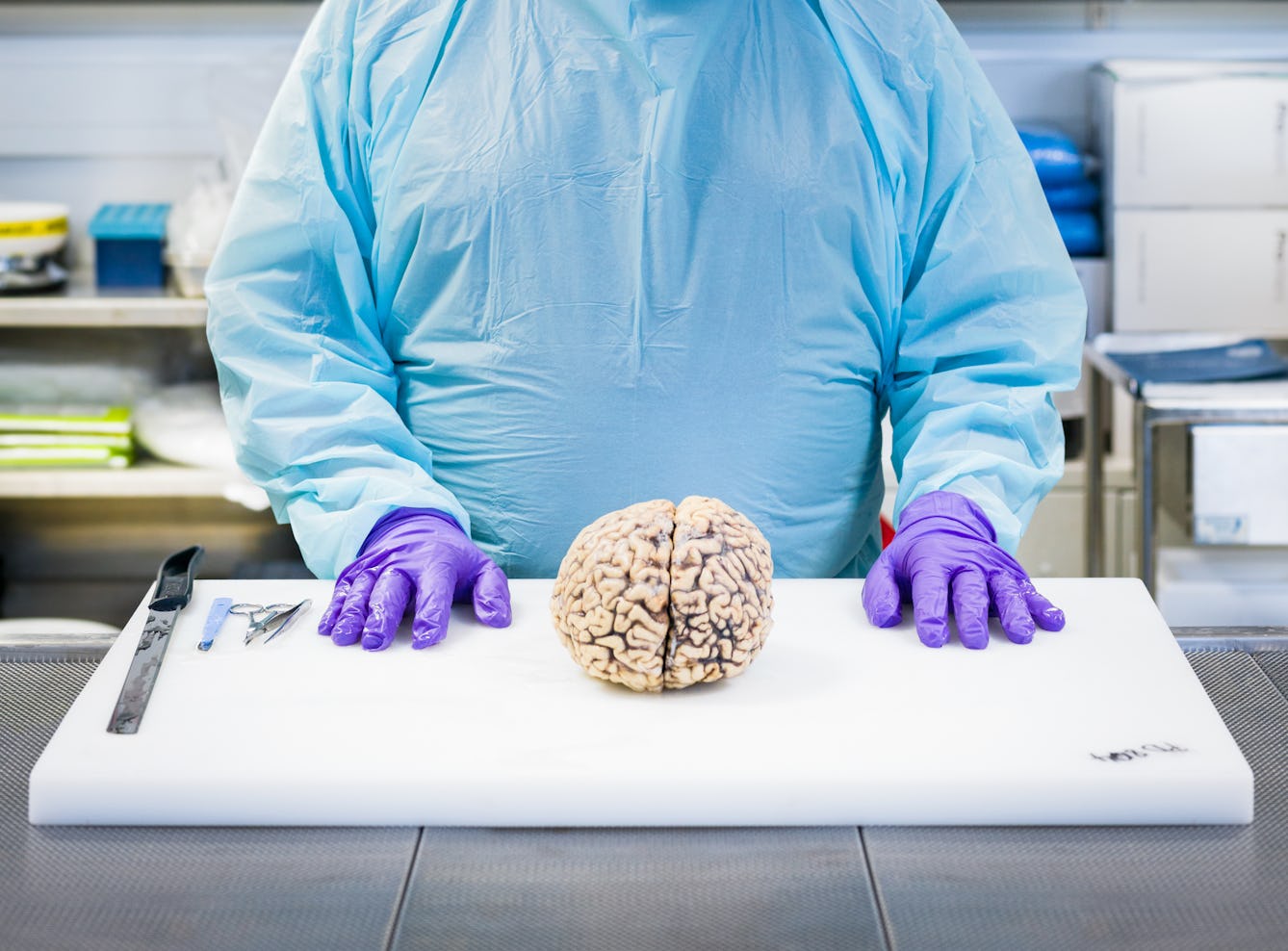
The anatomy of a brain dissection
Dissecting the brain after death not only helps confirm a diagnosis, but it can also teach us so much more about the symptoms and causes of brain diseases and how to treat them.

- Article
The solidarity of sickness
Visiting an injured friend in hospital prompts writer Sinéad Gleeson to reflect on the instant rapport forged between compatriots in the kingdom of the sick.
- Article
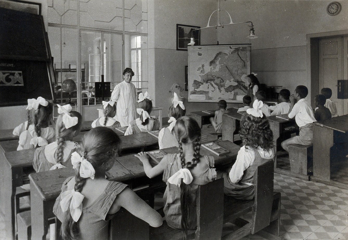
The law of periodicity for menstruation
Dr Edward Clarke's Law of Periodicity claimed that females who were educated alongside their male peers were developing their minds at the expense of their reproductive organs.
- Article
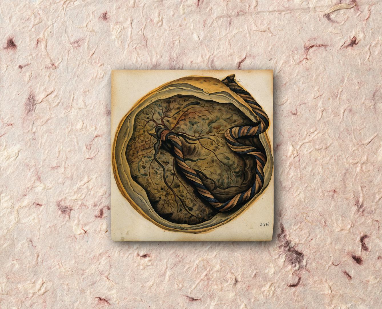
Womb milk and the puzzle of the placenta
A human baby needs milk to survive – and this holds true even before it’s born. Joanna Wolfarth explores “womb milk”, as well as ancient and modern ideas about the placenta.


Catalogue
- Archives and manuscripts
Pulteney, Richard (1730-1801), physician and botanist
Pulteney, Richard, 1730-1801.Date: 1759-1785Reference: MS.7441- Archives and manuscripts
Correspondence between Pulteney and Dr William Cuming
Date: 1779-1785Reference: MS.7441/8-10Part of: Pulteney, Richard (1730-1801), physician and botanist
- Digital Images
- Online
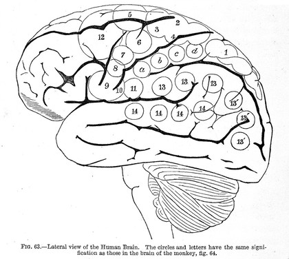
Addiction and reward pathways in the brain, artwork
Stephen Magrath
- Pictures
- Online
Brain: dissection showing cross-section through head and neck, with lateral view of the brain. Coloured line engraving by W.H. Lizars, ca. 1827.
Lizars, W. H. (William Home), 1788-1859.Date: [1820/1827]Reference: 563616i- Archives and manuscripts
Society of Medical Officers of Health
Society of Medical Officers of HealthDate: 1856-1998Reference: SA/SMO