Stories
- Article
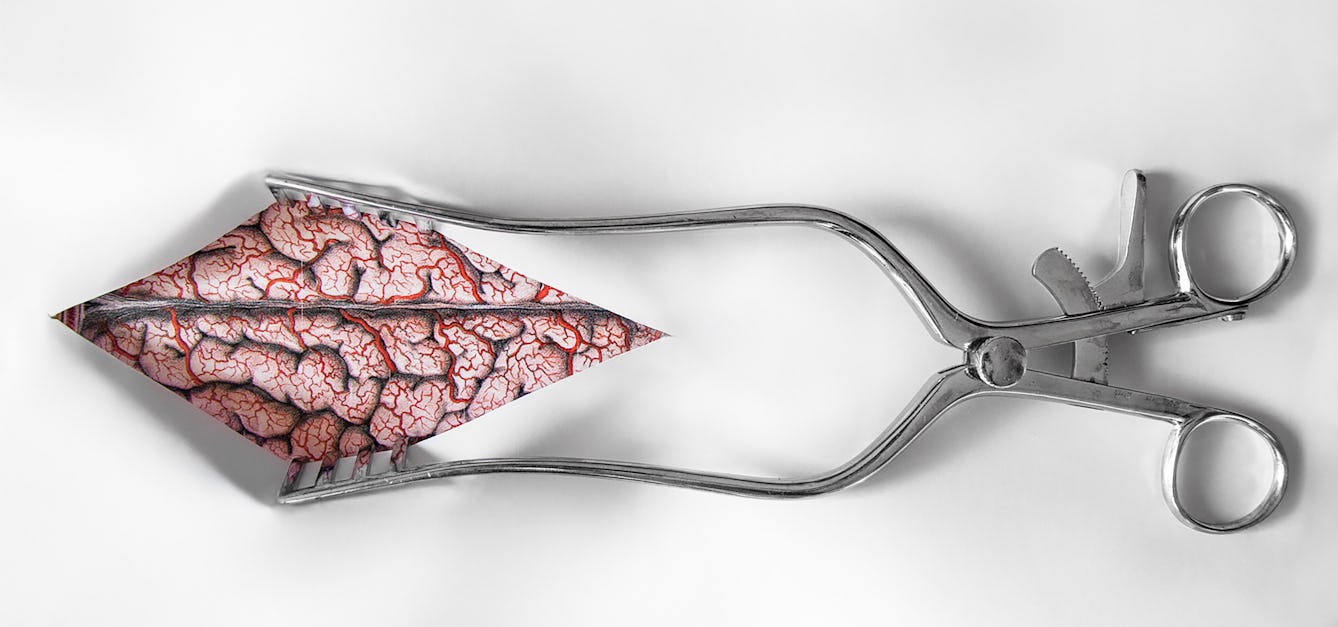
The mystery of the malignant brain
In 1884 a neurologist successfully used a patient’s symptoms, plus a new kind of map, to locate a brain tumour. Discover how his best-laid plans for treatment worked out.
- Article
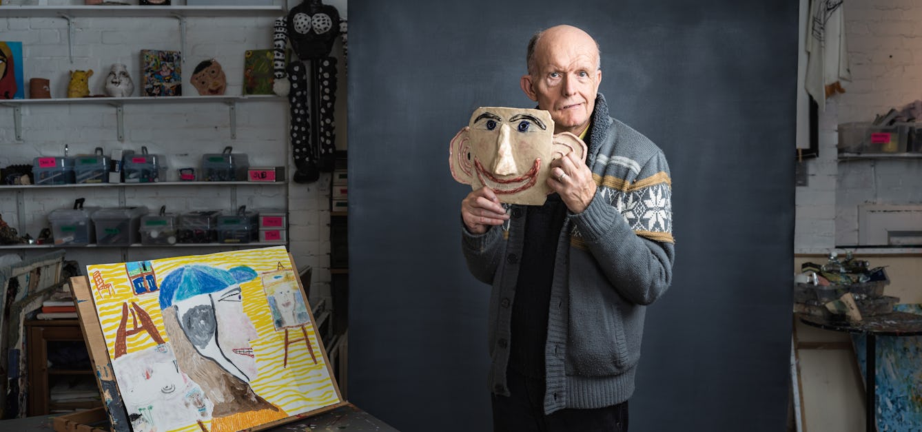
Rebuilding my identity after a brain injury
Chris Miller talks about how a brain injury forced him to reassess his place in the world – physically, personally and socially.

- Article
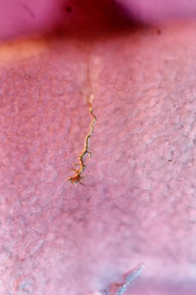
Cracks that let the light in
Rai Waddingham lives with voices other people cannot hear. Here she describes how she has come to accept, understand and calm her voices, and to acknowledge her strength.

- Article
The soul in the stomach
A 17th-century physician’s controversial theory about the link between the emotions and the stomach reminds us that we shouldn’t ignore our ‘gut feelings’.


Catalogue

- Digital Images
- Online
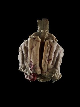
Brain with masses of new growth in the cerebrum
Godart, Thomas- Archives and manuscripts
'Chemistry of the Brain' and 'Data collected c.1950': ts and ms drafts re 'growth of biochemical knowledge of the brain'
Date: c.1950Reference: PP/MCI/D/8Part of: McIlwain, Henry- Archives and manuscripts
'Chemistry of the Brain' and 'Data collected c.1950': ts and ms drafts re 'growth of biochemical knowledge of the brain'
Date: c.1950Reference: PP/MCI/D/9Part of: McIlwain, Henry- Books
Brain repair / Donald G. Stein, Simón Brailowsky, Bruno Will.
Stein, Donald G.Date: 1997, ©1995- Videos
Brain doctors. Part 2, The Decision.
Date: 2013