37 results filtered with: Animal
- Digital Images
- Online
Arterial nodules, horse
Michael Frank, Royal Veterinary College
- Digital Images
- Online
Lungs and collapsed trachea, canine
Michael Frank, Royal Veterinary College
- Digital Images
- Online
3D reconstructed elephant hind limbs
Scott Birch
- Digital Images
- Online
Kidneys showing bilateral atrophy (tissue wasting)
Michael Frank, Royal Veterinary College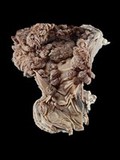
- Digital Images
- Online
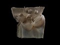
Cow foregut showing multiple warty growths (papillomas). These have grown from the gut lining, which is formed of squamous epithelium (consisting of flat, thin cells). These benign (non-cancerous) tumours can be caused by papillomavirus infection.
Michael Frank, Royal Veterinary College
- Digital Images
- Online
Microvasculature of the African Grey Parrot
Scott Birch, Scott Echols
- Digital Images
- Online
Canine lungs showing anthracosis (carbon accumulation)
Michael Frank, Royal Veterinary College
- Digital Images
- Online
Animal Materia medica.

- Digital Images
- Online
Dog (puppy) with cleft palate
Michael Frank, Royal Veterinary College
- Digital Images
- Online
Microvasculature of the African Grey Parrot
Scott Birch, Scott Echols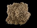
- Digital Images
- Online
Horse intestine with multiple attached parasitic worms
Michael Frank, Royal Veterinary College
- Digital Images
- Online
Canine skull with osteosarcoma
Michael Frank, Royal Veterinary College