419 results filtered with: Digital Images, Pictures

- Digital Images
- Online
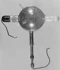
An X-ray valve tube, common type. Inscribed 'Made in England by Cuthbert Andrews'.
Cuthbert Andrews
- Pictures
- Online
X-ray of a rib-cage (?), showing matter (shrapnel ?). Photograph, ca. 1915.
Date: 1915Reference: 577685i
- Digital Images
- Online
X-ray tubes. Modified version of the Coolidge tube. G.R. Doglass insert tube (water-cooled) tube.
- Pictures
- Online
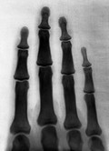
The bones of the human fingers. Photograph of X-ray attributed to L. Ropner, 1897.
Ropner, L., active 1897.Date: 2 November 1897Reference: 25612i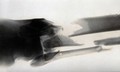
- Pictures
- Online
X-ray of a leg, showing broken bones (the tibula and fibia). Photograph, ca. 1915.
Date: 1915Reference: 577649i
- Digital Images
- Online
X-ray tube. Inscribed 'Cox's Record Regulating tube.' Suitable for treatment or radiograph with light currents.

- Digital Images
- Online
An X-ray valve tube, common in blue glass. Inscribed 'Made in England by Cuthbert Andrews'
Cuthbert Andrews
- Pictures
- Online
X-ray of a leg, showing a safety pin and a broken fibula. Photograph, ca. 1915.
Date: 1915Reference: 577652i
- Pictures
- Online
X-ray photograph of a skull, probably from a person with Down's syndrome. Photograph by Finzi, 1913.
Finzi, Neville Samuel, 1881-1968.Date: 1913Reference: 39115i
- Pictures
- Online
X-ray of a knee or elbow joint, showing matter (shrapnel ?) in the tissue. Photograph, ca. 1915.
Date: 1915Reference: 577661i- Pictures
Heart surgery: Denton A. Cooley and Domingo Liotta examining an X-ray of the first artificial heart. Photograph, 1969.
Date: 1969Reference: 583028i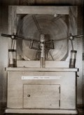
- Pictures
- Online
X-ray machine used by the physiologist Walter Bradford Cannon to investigate the mechanical process of digestion, 1896/1898. Colour photograph.
Date: 1900-1999Reference: 577232i
- Pictures
- Online
Surgeons examining a Mauser bullet in a man's chest via the use of an X-ray. Halftone, 1900, after W. Small.
Small, William, 1843-1929.Date: 1900Reference: 23558i
- Pictures
- Online
The bones of the wrist of Mrs Herries: two views. Photograph of X-ray by G. Harrison Orton, 1918.
Orton, G. Harrison (George Harrison), 1873-Date: 1918Reference: 662702i
- Pictures
- Online
X-ray (of an arm ?), showing pins in the bones (the radius and ulna ?). Photograph, ca. 1915.
Date: 1915Reference: 577650i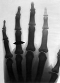
- Pictures
- Online
The bones of the human fingers, wearing a finger ring. Photograph of X-ray attributed to L. Ropner, 1897.
Ropner, L., active 1897.Date: 2 November 1897Reference: 25613i
- Digital Images
- Online
X-ray tube. Heavy and anti-cathode type. The anti cathode has a curious funnel-like projection facing the anode.
- Pictures
- Online
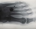
X-ray of a hand or foot, showing a (bullet hole ?) between the third and fourth digits. Photograph, ca. 1915.
Date: 1915Reference: 577654i
- Pictures
- Online
The bones of the hand of Mrs F. Bridgeman, wearing a finger ring, showing a broken wrist. Photograph of X-ray, 1911.
Date: 1911Reference: 662326i
- Pictures
- Online
Wilhelm Conrad Roentgen looking into an X-ray screen placed in front of a man's body and seeing the ribs and the bones of the arm. Chromolithograph.
Date: [1896/1900]Reference: 38591i
- Digital Images
- Online
An X-ray tube, common type. Inscribed 'Instanta' Tube - Macalister-Wiggin Pattern. Sole British Manufacturers Newton and Wright. Bears Ministry of Munitions Label.

- Digital Images
- Online
X-ray tube. Heavy anti-cathode (Gundelach type). The anti-cathode of tungsten is set in copper and is inscribed 'Patented Sept 5 1911'.
- Digital Images
- Online
An X-ray valve tube designed by Sir Oliver Lodge. Inscribed "Sir O. Lodge. Patent C 685". Made by Watson's 25.8.15. Made of Amber glass.

- Digital Images
- Online
Crystals of a DNA repair protein from Serratia marscescens bound to DNA. The crystals are grown in very small drops (approximatetly 1 microlitre) from very pure preparations so their structure can be determined by X-ray crystallography.
Bernard O'Hara & Renos Savva
- Digital Images
- Online
Crystals of a DNA repair protein from Serratia marscescens bound to DNA. The crystals are grown in very small drops (approximatetly 1 microlitre) from very pure preparations so their structure can be determined by X-ray crystallography.
Bernard O'Hara & Renos Savva