157 results filtered with: Pictures
- Pictures
X-ray of a female pelvis, showing a Grafenberg ring (intrauterine device) in position. X-ray, ca. 1945?.
Date: [1945?]Reference: 580006i- Pictures
X-ray of a female pelvis, showing a Grafenberg ring (intrauterine device) in position. X-ray, ca. 1945?.
Date: [1945?]Reference: 580007i- Pictures
X-ray of a female pelvis, showing a Grafenberg ring (intrauterine device) in position. X-ray, ca. 1945?.
Date: [1945?]Reference: 580010i- Pictures
X-ray of a hand with a red star, indicating the need of X-ray units in Russia. Colour photograph of a colour lithograph after A. Games, 1942.
Games, Abram, 1914-1996.Date: 1994Reference: 20292i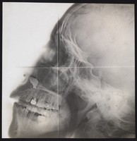
- Pictures
- Online
X-ray of a skull, in profile. Photograph, ca. 1915.
Date: 1915Reference: 577687i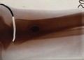
- Pictures
- Online
X-ray of a leg (shinbone ?). Photograph, ca. 1900.
Date: 1900Reference: 577226i
- Pictures
- Online
X-ray of a foot, showing a bullet. Photograph, ca. 1915.
Date: 1915Reference: 577664i
- Pictures
- Online
X-ray of a pair of hands with damaged fingers. Photograph, ca. 1915.
Date: 1915Reference: 577698i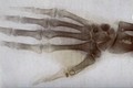
- Pictures
- Online
X-ray of a hand, showing damage to the thumb. Photograph, ca. 1915.
Date: 1915Reference: 577655i
- Pictures
- Online
X-ray (of an arm ?), showing a broken bone. Photograph, ca. 1915.
Date: 1915Reference: 577651i
- Pictures
- Online
X-ray of a rib-cage (?), showing matter (shrapnel ?). Photograph, ca. 1915.
Date: 1915Reference: 577685i
- Pictures
- Online
X-ray of a leg, showing a safety pin and a broken fibula. Photograph, ca. 1915.
Date: 1915Reference: 577652i
- Pictures
- Online
X-ray photograph of a skull, probably from a person with Down's syndrome. Photograph by Finzi, 1913.
Finzi, Neville Samuel, 1881-1968.Date: 1913Reference: 39115i
- Pictures
- Online
X-ray of a knee or elbow joint, showing matter (shrapnel ?) in the tissue. Photograph, ca. 1915.
Date: 1915Reference: 577661i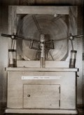
- Pictures
- Online
X-ray machine used by the physiologist Walter Bradford Cannon to investigate the mechanical process of digestion, 1896/1898. Colour photograph.
Date: 1900-1999Reference: 577232i
- Pictures
- Online
X-ray (of an arm ?), showing pins in the bones (the radius and ulna ?). Photograph, ca. 1915.
Date: 1915Reference: 577650i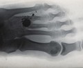
- Pictures
- Online
X-ray of a hand or foot, showing a (bullet hole ?) between the third and fourth digits. Photograph, ca. 1915.
Date: 1915Reference: 577654i
- Pictures
- Online
A deformed foot: X-ray. Photograph.
Date: 1900-1999Reference: 590650i
- Pictures
X-ray of a hand. Photograph, ca. 1900 (?).
Date: 1900Reference: 577235i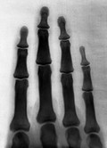
- Pictures
- Online
The bones of the human fingers. Photograph of X-ray attributed to L. Ropner, 1897.
Ropner, L., active 1897.Date: 2 November 1897Reference: 25612i
- Pictures
- Online
X-ray of a leg, showing broken bones (the tibula and fibia). Photograph, ca. 1915.
Date: 1915Reference: 577649i- Pictures
Heart surgery: Denton A. Cooley and Domingo Liotta examining an X-ray of the first artificial heart. Photograph, 1969.
Date: 1969Reference: 583028i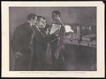
- Pictures
- Online
Surgeons examining a Mauser bullet in a man's chest via the use of an X-ray. Halftone, 1900, after W. Small.
Small, William, 1843-1929.Date: 1900Reference: 23558i
- Pictures
- Online
The bones of the wrist of Mrs Herries: two views. Photograph of X-ray by G. Harrison Orton, 1918.
Orton, G. Harrison (George Harrison), 1873-Date: 1918Reference: 662702i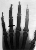
- Pictures
- Online
The bones of the human fingers, wearing a finger ring. Photograph of X-ray attributed to L. Ropner, 1897.
Ropner, L., active 1897.Date: 2 November 1897Reference: 25613i