86 results
- Pictures
Diseased adrenal glands, affected by tuberculosis and a struma, with a detail below showing cells, as seen under a microscope: three figures. Chromolithograph by W. Gummelt, ca. 1897.
Gummelt, W.Date: [1897?]Reference: 577694i
- Pictures
- Online
A diseased liver, including a section seen under a microscope, showing symptoms of atrophia hepatis acuta. Chromolithograph by W. Gummelt, ca. 1897.
Gummelt, W.Date: [1897?]Reference: 577041i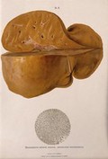
- Pictures
- Online
A diseased liver, including a section seen under a microscope, showing symptoms of degeneratio hepatis adiposa. Chromolithograph by W. Gummelt, ca. 1897.
Gummelt, W.Date: [1897?]Reference: 577033i
- Pictures
- Online
Dissections of diseased hearts: three figures, including hearts affected by forms of endocarditis and myocardial haemorrhage. Chromolithograph by W. Gummelt, ca. 1897.
Gummelt, W.Date: [1897?]Reference: 577233i
- Pictures
- Online
Dissections of diseased livers: four figures showing symptoms caused by hepatitis, syphilis, anfioma cavernosum and pylephlebitis. Chromolithograph by W. Gummelt, ca. 1897.
Gummelt, W.Date: [1897?]Reference: 577054i
- Pictures
- Online
Dissections of femoral arteries affected by calcification, with illustrations of subperitoneal phlebectasia and rectal rectal hemorrhoids. Chromolithograph by W. Gummelt, ca. 1897.
Gummelt, W.Date: [1897?]Reference: 577242i
- Pictures
- Online
Section through part of a tibia bone, showing diseased tissue and a cancerous (?) sarcoma (bone tumour). Chromolithograph by W. Gummelt, ca. 1897.
Gummelt, W.Date: [1897?]Reference: 577412i
- Pictures
- Online
Sections through two femur bones: the left illustration indicating the red bone marrow, the right showing the marrow and lymphatics. Chromolithograph by W. Gummelt, ca. 1897.
Gummelt, W.Date: [1897?]Reference: 577399i
- Pictures
- Online
Dissections of an ulcerated stomach caused by syphilis, and a severe case of ulcerated proctitis of the rectum (?): two figures. Chromolithograph by W. Gummelt, ca. 1897.
Gummelt, W.Date: [1897?]Reference: 577375i
- Pictures
- Online
Underside of a brain, shown beside a dissection of the abdominal aorta and left iliac artery, both showing symptoms of atherosclerosis and aneurysm. Chromolithograph by W. Gummelt, ca. 1897.
Gummelt, W.Date: [1897?]Reference: 577230i
- Pictures
- Online
Sections through humerus and femur bones, two figures: the left illustration indicating a sarcoma (bone tumour) on the humerus bone, the right showing cancerous (?) metastases in the marrow of the femur. Chromolithograph by W. Gummelt, ca. 1897.
Gummelt, W.Date: [1897?]Reference: 577408i