172 results
- Pictures
Discoloured, swollen left breast showing cancerous tumour, broken down and ulcerated in a 54-year old woman with pulmonary embolus and thrombosis. Watercolour by Barbara E. Nicholson, 1948.
Nicholson, BarbaraDate: 1948Reference: 32681iPart of: Barbara Nicholson medical illustration collection.- Archives and manuscripts
Box 5 Robert Macintosh in USSR and on Trans-Siberian Express, 1959, Japan 1960, Pask life jackets and resuscitation experiment 1942, boy with swollen face in Uganda 1962
Date: 1942-1962Reference: PP/RRM/F.47/147-176Part of: Macintosh, Sir Robert (1897-1989)
- Pictures
- Online
The severely diseased and swollen scrotum and legs of a man, with details showing diseased areas of skin on the leg and shoulder. Watercolour by C. D'Alton, 1840/1870?.
D'Alton, Christopher, active 1847-1871.Date: [1840/1870?]Reference: 574940i
- Pictures
- Online
George, Prince Regent, in uniform holding out a swollen hand which is supported by Wellington; representing the enormous amount of money given to the army compared with the navy. Coloured lithograph, 1816.
Date: [August 1816?]Reference: 12213i- Pictures
Oedema of larynx in a male patient: sketch of swollen neck and head showing sacs of excess accumulation of serous fluid. Pen and ink by Barbara E. Nicholson, 1958.
Nicholson, BarbaraDate: 1958Reference: 35939iPart of: Barbara Nicholson medical illustration collection.
- Pictures
- Online
Initial dissection of the pregnant female abdomen, showing the skin peeled away to reveal the swollen uterus. Copperplate engraving by F.S. Ravenet after I.V. Rymsdyk, 1774, reprinted 1851.
Rymsdyk, Jan van, active 1750-1788.Date: [1851]Reference: 579790iPart of: Hunter, William
- Pictures
- Online
Uganda: a Lango woman with diseased skin holding a baby; she has applied a leaf as a dressing to her swollen left breast. Photograph by Cecil John Hackett, ca. 1937.
Hackett, Cecil John, 1905-1995.Date: 1937Reference: 580450i- Pictures
Progressive migrating tumours, presented as swollen manifestation in the left lower jaw, in a 48-year old man with silent hypernephroma of the left kidney. Watercolour by Barbara E. Nicholson, 1947.
Nicholson, BarbaraDate: 1947Reference: 32130iPart of: Barbara Nicholson medical illustration collection.
- Pictures
- Online
A surgeon bleeding Ragotin's arm - upon waking and attempting to get dressed he discovered his clothes were too tight and he believes his body has swollen in the night. Engraving.
Reference: 22994i
- Pictures
- Online
Front view of the pregnant uterus and pelvic area, showing the skin peeled away to reveal the swollen womb. Copperplate engraving by R. Strange after I.V. Rymsdyk, 1774, reprinted 1851.
Rymsdyk, Jan van, active 1750-1788.Date: [1851]Reference: 579795iPart of: Hunter, William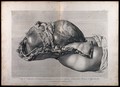
- Pictures
- Online
Dissection of the pregnant female abdomen, showing the skin peeled away to reveal the swollen uterus and the viscera, side view. Copperplate engraving by G. Scotin after I.V. Rymsdyk, 1774, reprinted 1851.
Rymsdyk, Jan van, active 1750-1788.Date: [1851]Reference: 579792iPart of: Hunter, William- Pictures
Fatal testicular cancer in a 66-year old man with secondaries in adrenals and kidney: swollen right testicle section showing tumour and surrounding growth in complete replacement. Watercolour by Barbara E. Nicholson, 1958.
Nicholson, BarbaraDate: 1958Reference: 36084iPart of: Barbara Nicholson medical illustration collection.
- Digital Images
- Online
Drawing of the 1918 Influenza: Trachea showing earliest stage of reaction in influenza. Epithelium intact, but basement [?] slightly swollen as [?], with congestion and some small cells influtration of sub[?]. Hyaline deposits over epithelium
John George Adami
- Pictures
- Online
A surgeon performing a paracentesis on an obese man, whose swollen abdomen has a cannula inserted into it, and is subsequently releasing fluid into a basin. Pen drawing by Z.S. after an engraving, 1672.
Reference: 22784i
- Pictures
- Online
Dissection of the pregnant female abdomen, showing the skin peeled away to reveal the swollen uterus, the diaphragm and the intestines, side view. Copperplate engraving by T. Major after I.V. Rymsdyk, 1774, reprinted 1851.
Rymsdyk, Jan van, active 1750-1788.Date: [1851]Reference: 579793iPart of: Hunter, William
- Pictures
- Online
A baby boy with a scarred and swollen face with a warning in spanish about the risk of HIV mothers transmitting the AIDS disease to babies; a poster from the America responds to Aids advertising campaign. Lithograph.
Date: [between 1990 and 1999]Reference: 667380i- Pictures
Schonlein's purpura in a 50-year old woman: sketch of (a) legs, anterior view and (b) tongue, showing swollen joints and discolouration (resulting from extravasation of blood into the skin and mucus membranes). Watercolour by Barbara E. Nicholson, 1951.
Nicholson, BarbaraDate: 1951Reference: 34396iPart of: Barbara Nicholson medical illustration collection.- Pictures
Schoenlein's purpura in a 50-year old woman: sketch of (a) buttocks and legs and (b) hands, anterior and posterior views, showing swollen joints and discolouration (resulting from extravasation of blood into the skin and mucus membranes). Watercolour by Barbara E. Nicholson, 1951.
Nicholson, BarbaraDate: 1951Reference: 34320iPart of: Barbara Nicholson medical illustration collection.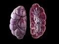
- Digital Images
- Online
Acute nephritis in calf kidneys
Michael Frank, Royal Veterinary College
- Digital Images
- Online
C18 Chinese woodcut: One-sided pharyngitis
- Digital Images
- Online
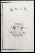
C18 Chinese woodcut: Bright red throat abscess
- Film
Proteus (bacteriaceae) : restricted forms as a result of the effect of penicillin.
Date: 1959
- Digital Images
- Online
C18 Chinese woodcut: Galloping pharyngitis

- Digital Images
- Online
Fucus Vesiculosus (Bladderwrack)
Rowan McOnegal
- Digital Images
- Online
Woman, possibly with a goitre
St Bartholomew's Hospital Archives & Museum