172 results
- Pictures
Section of a swollen thyroid gland, in a female patient. Watercolour by Barbara E. Nicholson, 1946.
Nicholson, BarbaraDate: 1946Reference: 31829iPart of: Barbara Nicholson medical illustration collection.
- Pictures
- Online
Diseased, swollen fingertip, with bone shown protruding from the open wound. Watercolour by C. D'Alton, 1856.
D'Alton, Christopher, active 1847-1871.Date: 1856Reference: 574459i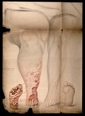
- Pictures
- Online
The swollen legs and the damaged feet of a woman with disease diagnosed as syphilis. Watercolour by C. D'Alton, 1868.
D'Alton, Christopher, active 1847-1871.Date: Nov. 10" 1868Reference: 730574i
- Pictures
- Online
A swollen and inflamed foot: gout is represented by an attacking demon. Etching, 1835, after J. Gillray, 1799.
Gillray, James, 1756-1815.Date: 1 August 1835Reference: 10508i
- Books
- Online
On retrobulbar incision of the sheath of the optic nerve in cases of swollen disc / by Robert Brudenell Carter.
Carter, Robert Brudenell, 1828-1918.Date: 1887
- Pictures
- Online
A swollen and inflamed foot: gout is represented as an attacking demon. Coloured etching, 1835, after J. Gillray, 1799.
Gillray, James, 1756-1815.Date: 1 August 1835Reference: 10509i
- Pictures
- Online
Five demons: one performs dental surgery on another, while another with a swollen mouth waits for attention. Coloured etching.
Reference: 11643i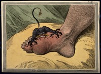
- Pictures
- Online
A swollen and inflamed foot: gout is represented by an attacking demon. Coloured soft-ground etching by J. Gillray, 1799.
Gillray, James, 1756-1815.Date: 14 May 1799Reference: 10507i- Pictures
Blood flow of a swollen, discoloured left foot in a female patient with gangrene. Watercolour by Barbara E. Nicholson, 1947.
Nicholson, BarbaraDate: 1947Reference: 31993iPart of: Barbara Nicholson medical illustration collection.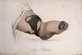
- Pictures
- Online
A swollen foot and ankle with a skin disease. Coloured stipple engraving by S. Tresca after Moreau-Valvile, c. 1806.
Moreau-Valvile, active 1806.Reference: 29681i
- Pictures
A naked man, standing; despite his emaciation, his abdomen is swollen. Photograph by L. Haase after H.W. Berend, 1864.
Berend, H. W. (Heimann Wolff), 1809-1873.Date: 1856Reference: 31956i- Pictures
Urine reflux in a male patient with swollen testis: specimen showing thickened, congested epididymis. Watercolour by Barbara E. Nicholson, 1956.
Nicholson, BarbaraDate: 1956Reference: 35718iPart of: Barbara Nicholson medical illustration collection.
- Pictures
- Online
Two examples of a diseased penis: the first with a grossly swollen glans and the second excreting pus. Chromolithograph, c. 1888.
Reference: 35072i- Books
Case of swollen optical disc, in which the sheath of the optic nerve was incised behind the eyeball / by Robert Brudenell Carter.
Carter, Richard B. (Richard Burnett), 1931-Date: 1887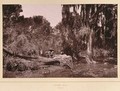
- Pictures
- Online
Lover's Walk, South Africa: a narrow lane obstructed by an uprooted tree and swollen stream. Woodburytype, 1888, after a photograph by Robert Harris.
Harris, Robert, active 1881-1888.Date: 1888Reference: 533305iPart of: Harris, Robert, fl. 1881/1888.- Pictures
Cervical polyp in a 55-year old woman: surgical specimen and section showing swollen hypertrophied tumerous membrane. Watercolour by Barbara E. Nicholson, 1951.
Nicholson, BarbaraDate: 1951Reference: 34314iPart of: Barbara Nicholson medical illustration collection.
- Digital Images
- Online
Lover's Walk, South Africa: a narrow lane obstructed by an uprooted tree and swollen stream. Woodburytype, 1888, after a photograph by Robert Harris.
Robert Harris
- Digital Images
- Online
(Left) Representing the leg swollen and ulcerated from the effects of Secondary symptoms. (Right) Representing the face covered with Tubercles & veneral excrescences

- Pictures
- Online
Uganda: the swollen and bandaged toes of a member of the Lango tribe, possibly suffering from yaws. Photograph by Cecil John Hackett, ca. 1937.
Hackett, Cecil John, 1905-1995.Date: 1937Reference: 580331i- Pictures
Familial polyposis in a male patient: subtotal colectomy specimen showing polypoid projection of swollen, hypertrophied and tumorous membrane. Watercolour by Barbara E. Nicholson, 1956.
Nicholson, BarbaraDate: 1956Reference: 35547iPart of: Barbara Nicholson medical illustration collection.- Pictures
Acute appendicitis in a 44-year old man: specimen detail from paracolic gutter showing inflammatory changes and hard swollen tip. Watercolour by Barbara E. Nicholson, 1953.
Nicholson, BarbaraDate: 1953Reference: 35013iPart of: Barbara Nicholson medical illustration collection.
- Pictures
- Online
A child with a swollen mouth being led by a woman to visit an apothecary, who is seated in his laboratory. Drawing by William Heath Robinson.
Robinson, W. Heath (William Heath), 1872-1944.Reference: 39058i- Pictures
A Chinese physician (?) pointing to the swollen abdomen of a pregnant (?) woman, who is reclining on a bed. Pen and ink with watercolour, China, 18--.
Date: 1800-1899Reference: 567483iPart of: Various diseases found in Chinese men and women.
- Pictures
- Online
A swollen lethargic patient with ten physicians seated around a table on which are axes and a halter; symbolising England's government and the need for reform. Engraving, 1756.
Date: 23 July 1756Reference: 10760i- Pictures
Diseased colon in a 43-year old man with cancer of rectum: showing projecting fungus-like mass of swollen, hypertrophied, tumorous membrane. Watercolour by Barbara E. Nicholson, 1949.
Nicholson, BarbaraDate: 1949Reference: 33019iPart of: Barbara Nicholson medical illustration collection.