312 results

- Pictures
- Online
Female genitalia held open by two fingers to show an area of diseased tissue. Watercolour by Christopher D' Alton.
D'Alton, Christopher, active 1847-1871.Date: 1800-1899Reference: 38124i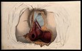
- Pictures
- Online
The female reproductive system: dissection of the genitalia and anus. Coloured line engraving by W.H. Lizars, ca. 1827.
Lizars, W. H. (William Home), 1788-1859.Date: [1827?]Reference: 579935i
- Pictures
- Online
Female genitalia showing severely diseased tissue spreading on to the right inside thigh. Watercolour by Christopher D' Alton, 1865.
D'Alton, Christopher, active 1847-1871.Date: 1865Reference: 38295i
- Pictures
- Online
Female genitalia held open by a finger to show an area of diseased tissue. Watercolour by Christopher D' Alton.
D'Alton, Christopher, active 1847-1871.Date: 1800-1899Reference: 38132i
- Pictures
- Online
Female genitalia held open by two fingers to show an area of diseased tissue. Watercolour by C. D'Alton, 18--.
D'Alton, Christopher, active 1847-1871.Date: 1800-1899Reference: 573917i
- Pictures
- Online
Female genitalia held open by two fingers to show an area of diseased tissue. Watercolour by Christopher D' Alton, 1857.
D'Alton, Christopher, active 1847-1871.Date: 1857Reference: 38103i
- Pictures
- Online
Female genitalia held open by two fingers to show an area of diseased tissue. Watercolour by Christopher D' Alton, 1866.
D'Alton, Christopher, active 1847-1871.Date: 1866Reference: 38137i
- Pictures
- Online
Female genitalia showing severely diseased tissue of the labia, and some sores on the thighs. Watercolour by Christopher D' Alton.
D'Alton, Christopher, active 1847-1871.Date: 1800-1899Reference: 38308i
- Pictures
- Online
Female genitalia held open by a finger to show an area of diseased tissue. Watercolour by Christopher D' Alton, 1857.
D'Alton, Christopher, active 1847-1871.Date: 1857Reference: 38106i
- Pictures
- Online
Female genitalia held open by the fingers to show an area of diseased tissue. Watercolour by Christopher D' Alton, 1857.
D'Alton, Christopher, active 1847-1871.Date: 1857Reference: 38133i
- Pictures
- Online
The female reproductive system, showing an external view of the genitalia. Coloured line engraving by W.H. Lizars, ca. 1827.
Lizars, W. H. (William Home), 1788-1859.Date: [1827?]Reference: 579934i
- Pictures
- Online
Female genitalia held open by two fingers to show an area of diseased tissue. Watercolour by Christopher D' Alton, 1857.
D'Alton, Christopher, active 1847-1871.Date: 1857Reference: 38056i
- Pictures
- Online
Female genitalia held open by a finger to show an area of diseased tissue. Watercolour by Christopher D' Alton, 1866.
D'Alton, Christopher, active 1847-1871.Date: [18]66Reference: 38130i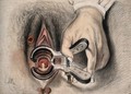
- Pictures
- Online
An infected sore on the female genitalia shown with the aid of a vaginal speculum. Watercolour by C.D' Alton, 18--.
D'Alton, Christopher, active 1847-1871.Date: 1800-1899Reference: 573913i
- Pictures
- Online
Female genitalia showing severely diseased tissue, and sores on the upper thighs to each side. Watercolour by Christopher D' Alton, 1857.
D'Alton, Christopher, active 1847-1871.Date: 1857Reference: 38166i
- Pictures
- Online
Female genitalia showing severely diseased tissue, and sores on the inside thighs, buttocks and pubis. Watercolour by Christopher D' Alton, 1856.
D'Alton, Christopher, active 1847-1871.Date: 1856Reference: 38196i
- Books
- Online
Gynecological pathology : a manual of microscopic technique and diagnosis in gynecological practice, for students and physicians / by Dr. Carl Abel, tr. and ed. by Samuel Wyllis Bandler. With a chapter on the embryology of the female genitalia and the pathological growths developing from embryonal structures. Illustrated by one hundred engravings.
Abel, Karl, 1863-Date: 1901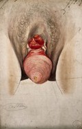
- Pictures
- Online
Female genitalia showing severely diseased tissue and symptoms of ectropia vesicae, with a complete prolapse of the uterus. Watercolour by Christopher D'Alton, ca. 1847.
D'Alton, Christopher, active 1847-1871.Date: [1847?]Reference: 573954i
- Pictures
- Online
Female genitalia showing severely diseased tissue from pubis to anus, and a large growth on the left inside thigh. Watercolour by Christopher D' Alton, 1858.
D'Alton, Christopher, active 1847-1871.Date: 1858Reference: 38274i
- Pictures
- Online
Female genitalia with an area of diseased tissue: a surgical instrument is shown extracting part of the diseased area. Watercolour by C. D' Alton, 1858.
D'Alton, Christopher, active 1847-1871.Date: 1858Reference: 573924i
- Pictures
- Online
Female genitalia with areas of diseased tissue, and clusters of sores on the top of the thighs to each side. Watercolour by Christopher D' Alton, 1856.
D'Alton, Christopher, active 1847-1871.Date: 1856Reference: 38190i- Books
Notes on some Indian species of the genus Phlebotomus. Part XIX, The value of the female genitalia in the identification of species / by J.A. Sinton.
Sinton, J. A. (John Alexander), 1884-1956.Date: 1927
- Pictures
- Online
A penis with a patch of skin disease on the shaft; and female genitalia with a skin disease around the labia and vulva. Chromolithograph, c. 1888.
Reference: 34905i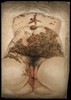
- Pictures
- Online
Female genitalia showing severely diseased tissue caused by syphilis: extensive sores and abcesses are seen extending up the abdomen and torso. Watercolour by C. D'Alton, 1862.
D'Alton, Christopher, active 1847-1871.Date: 1862Reference: 574404i
- Pictures
- Online
Female genitalia with sores around the anus: with details showing a sore on the genitals and a rash on a woman's chest. Watercolour by C. D'Alton, 1869.
D'Alton, Christopher, active 1847-1871.Date: [18]69Reference: 573923i