698 results
- Pictures
- Online
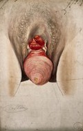
Female genitalia showing severely diseased tissue and symptoms of ectropia vesicae, with a complete prolapse of the uterus. Watercolour by Christopher D'Alton, ca. 1847.
D'Alton, Christopher, active 1847-1871.Date: [1847?]Reference: 573954i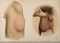
- Pictures
- Online
A section of leg with a swelling on the shin below the knee; and male genitalia with a lump on the testicles. Chromolithograph, c. 1888.
Reference: 34829i
- Pictures
- Online
Dissections of the male genitalia and upper thighs: four figures, with the arteries and blood vessels indicated in red. Coloured lithograph by J. Roux, 1822.
Roux, Jacob Chr.Date: [1822]Reference: 579776iPart of: Tiedemann, Friedrich, 1781-1861.
- Pictures
- Online
Female genitalia showing severely diseased tissue from pubis to anus, and a large growth on the left inside thigh. Watercolour by Christopher D' Alton, 1858.
D'Alton, Christopher, active 1847-1871.Date: 1858Reference: 38274i
- Pictures
- Online
Female genitalia with an area of diseased tissue: a surgical instrument is shown extracting part of the diseased area. Watercolour by C. D' Alton, 1858.
D'Alton, Christopher, active 1847-1871.Date: 1858Reference: 573924i
- Pictures
- Online
Diseased skin surrounding the anus and genitalia of a woman, with a detail of the area after treatment (?) Watercolour by C. D'Alton, ca. 1858.
D'Alton, Christopher, active 1847-1871.Date: [1858?]Reference: 574405i
- Pictures
- Online
Dissection of the area surrounding the anus and genitalia of a man, with the muscles and blood vessels indicated. Colour lithograph by G.H. Ford, 1865.
Ford, G. H. (George Henry)Date: 1865Reference: 580349i
- Pictures
- Online
Dissection of the area surrounding the anus and genitalia of a man, with the muscles and blood vessels indicated. Colour lithograph by G.H. Ford, 1865.
Ford, G. H. (George Henry)Date: 1865Reference: 580356i
- Pictures
- Online
Female genitalia with areas of diseased tissue, and clusters of sores on the top of the thighs to each side. Watercolour by Christopher D' Alton, 1856.
D'Alton, Christopher, active 1847-1871.Date: 1856Reference: 38190i- Books
Notes on some Indian species of the genus Phlebotomus. Part XIX, The value of the female genitalia in the identification of species / by J.A. Sinton.
Sinton, J. A. (John Alexander), 1884-1956.Date: 1927
- Pictures
- Online
A penis with a patch of skin disease on the shaft; and female genitalia with a skin disease around the labia and vulva. Chromolithograph, c. 1888.
Reference: 34905i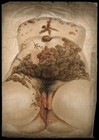
- Pictures
- Online
Female genitalia showing severely diseased tissue caused by syphilis: extensive sores and abcesses are seen extending up the abdomen and torso. Watercolour by C. D'Alton, 1862.
D'Alton, Christopher, active 1847-1871.Date: 1862Reference: 574404i
- Pictures
- Online
Female genitalia with sores around the anus: with details showing a sore on the genitals and a rash on a woman's chest. Watercolour by C. D'Alton, 1869.
D'Alton, Christopher, active 1847-1871.Date: [18]69Reference: 573923i
- Pictures
- Online
An écorché figure seen from behind, with details of the muscles of the hand and foot, and the male genitalia: five figures. Line engraving by A. Bell, 1788/1797.
Bell, Andrew, 1726-1809.Date: [1788/1797]Reference: 562356i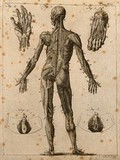
- Pictures
- Online
An écorché figure seen from behind, with details of the muscles of the hand and foot, and the male genitalia: five figures. Line engraving by A. Bell, 1771/1783.
Bell, Andrew, 1726-1809.Date: [1771/1783?]Reference: 562254i
- Pictures
- Online
Male genitalia with a skin disease; and varieties of sores which appeared on other parts of the man's body. Coloured aquatint by Bennett after J. Harrison, c. 1820.
Harrison, John.Date: c.1820Reference: 29842i
- Books
- Online
Gynecological pathology : a manual of microscopic technique and diagnosis in gynecological practice, for students and physicians / by Dr. Carl Abel, tr. and ed. by Samuel Wyllis Bandler. With a chapter on the embryology of the female genitalia and the pathological growths developing from embryonal structures. Illustrated by one hundred engravings.
Abel, Karl, 1863-Date: 1901
- Pictures
- Online
Female genitalia and uterus during pregnancy: six figures, including cross-sections of foetus and twins in utero. Line engraving, 1790(?), by W. Taylor after F. Birnie after W. Smellie.
Smellie, William, 1697-1763.Date: [1790?]Reference: 561818i
- Pictures
- Online
Female genitalia held open by the hand to show diseased tissue, with clusters of sores at the top of the thighs to each side. Watercolour by Christopher D' Alton, 1857.
D'Alton, Christopher, active 1847-1871.Date: 1857Reference: 38112i
- Books
- Online
Quaestio medica, quodlibetariis disputationibus manè discutienda in scholis medicorum, die Martis qnarto [sic] mensis Decembris, anno Domini M.DCC.LXX. M. Josepho-Ignatio Guillotin, doctore medico, praeside. An praeter genitalia sexus inter se discrepent?
Guillotin, Joseph Ignace, 1738-1814Date: [1770]- Pictures
The lower part of a naked man's body in colour with his penis in black and white; promoting consideration of a person beyond their genitalia, with implications for AIDS transmission. Colour lithograph after M. Kautter, ca. 2003.
Date: [2003?]Reference: 723677i
- Pictures
- Online
Three packets of blue condoms in the shape of male genitalia with the message 'progress or not, one still dies of AIDS'; advertising safe sex. Colour lithograph by Comed for the Ministère de la Santé, Division de la Médicine Préventive, Luxembourg.
Date: [between 1990 and 1999]Reference: 672122i- Archives and manuscripts
Correspondence about 2 brothers excruciating pains in arms etc
Date: 1955Reference: PP/FPW/A.5/10/2Part of: Parkes Weber, Frederick (1863-1962)
- Books
- Online
Bakteriologie des weiblichen Genitalkanales / von C. Menge und B. Krönig.
Menge Carl.Date: 1897
- Books
- Online
Contributo alla conoscenza delle terminazioni nervose negli organi genitali esterni e nel capezzolo della femmina / [Pasquale Sfameni].
Sfameni, Pasquale.Date: 1901