367 results filtered with: Pictures
- Pictures
- Online
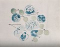
Cells, as seen under a microscope. Watercolour ca. 1912.
Date: [1912?]Reference: 576961i
- Pictures
- Online
Cells and organisms, as seen through a microscope: twenty figures. Lithograph by L. Aldous, 1846.
Aldous, Lens.Date: [1846]Reference: 576547i- Pictures
Germinal matter: eighteen figures showing granules, globules, fibres and cells. Line illustration, 1900/1940?.
Date: [1900/1940?]Reference: 569556i- Pictures
Shigellosis: acute: microscopic view of macrophage cells (white blood cells). Drawing by P.H. Manson-Bahr, ca. 1930.
Manson-Bahr, Philip H. (Philip Henry), Sir, 1881-1966.Date: 1930Reference: 571049i- Pictures
Germinal matter: eight figures including cells from humans, a boa and a dog. Line illustration, 1900/1940?.
Date: [1900/1940?]Reference: 569558i- Pictures
Germinal matter: ten figures including cartilage, tendon, nerve, muscle and pus cells of frogs and cats. Line illustration, 1900/1940?.
Date: [1900/1940?]Reference: 569572i
- Pictures
- Online
Microscope drawings of plant cells in glass tubes. Coloured lithograph by C. Varley, c.1834.
Varley, Cornelius, 1781-1873.Date: 1834Reference: 20414i- Pictures
Germinal matter: ten figures including brain, tooth, cornea, fat and starch-holding cells from humans, potatoes, frogs and salamanders. Line illustration, 1900/1940?.
Date: [1900/1940?]Reference: 569568i- Pictures
Nerve cells (?). Colour lithograph by F. Reichhold, 1895.
Reichhold, F.Date: [1895]Reference: 569159i
- Pictures
- Online
Microscope drawings of plant anatomy with cells and growing points. Lithograph after C. Varley, c.1834.
Varley, Cornelius, 1781-1873.Date: 1834Reference: 20432i- Pictures
Urea and uric acid crystals: nine figures, also showing crystals of hippuric acid, taurin, and leucine, with epithelial and cholestrine cells and myelin particles. Line illustration, 1900/1940?.
Date: [1900/1940?]Reference: 569588i- Pictures
Nerve cells (?) and the brain: three figures, including two diagrams showing sections through the brain, and one illustrating nerve cells (?). Colour lithograph by F. Reichhold, 1895.
Reichhold, F.Date: [1895]Reference: 569035i- Pictures
Nerve cells (?): six figures. Colour lithograph by F. Reichhold, 1895.
Reichhold, F.Date: [1895]Reference: 569161i- Pictures
Microscopy: cells, and parts of a plant. Engraving [after R. Hooke?].
Hooke, Robert, 1635-1703.Reference: 46513i
- Pictures
- Online
Microscope drawings of plant anatomy with cells and tissues of stonewort (Chara and Nitella species). Lithograph after C. Varley, c.1834.
Varley, Cornelius, 1781-1873.Date: 1834Reference: 20434i
- Pictures
- Online
A head divided into 35 cells representing human faculties. Woodcut, 1888, after M. Mihara.
Mihara, Muneaki, active ca. 1888.Date: Meiji 21 [1888]Reference: 2829831i- Pictures
Nerve cells (?) and the spinal cord: five figures, including nerve cells as seen through a microscope, and a cross section of the spinal cord. Wood engraving by M. Toller (?), 1895.
Toller, M.Date: [1895]Reference: 569232i- Pictures
Middlesex House of Correction: plan showing the cells, courtyards, colonnades, and dayrooms. Engraving 1821.
Sibley, Robert, active 1828.Date: [1821]Reference: 585001i- Pictures
Bone marrow cells on agar plate: scopic view. Watercolour by Barbara E. Nicholson, 1958.
Nicholson, BarbaraDate: 1958Reference: 36335iPart of: Barbara Nicholson medical illustration collection.- Pictures
Bone marrow cells on agar plate: scopic view. Watercolour by Barbara E. Nicholson, 1958.
Nicholson, BarbaraDate: 1958Reference: 36328iPart of: Barbara Nicholson medical illustration collection.- Pictures
Bone marrow cells on agar plate: scopic view. Watercolour by Barbara E. Nicholson, 1958.
Nicholson, BarbaraDate: 1958Reference: 36334iPart of: Barbara Nicholson medical illustration collection.
- Pictures
- Online
Brain and nerve cells in their healthy state and after injury by alcohol. Colour lithograph, ca. 1920.
Date: Reprinted 1930Reference: 679926iPart of: Why America went dry.
- Pictures
- Online
Two enlarged images of T-cells one infected with HIV by the Frederick Cancer Research and Development Center. Colour lithograph by Nancy Burson and Kunio Nagashima, 1991.
Date: 1991Reference: 667046i- Pictures
Nerve cells (?): three figures, as seen through a microscope. Colour lithograph by F. Reichhold, 1895.
Reichhold, F.Date: [1895]Reference: 569031i- Pictures
Pathological samples showing cells (possibly syphilis), as seen under a microscope. Watercolour by C. D'Alton, 18--.
D'Alton, Christopher, active 1847-1871.Date: [between 1800 and 1899]Reference: 575870i