168 results filtered with: Pictures
- Pictures
Bladder covered in cancerous tumours situated within the pelvis of a 46-year old man with haematuria and dysuria. Watercolour after radiograph by Barbara E. Nicholson, 1946.
Nicholson, BarbaraDate: 1946Reference: 31312iPart of: Barbara Nicholson medical illustration collection.- Pictures
A man gets up from his office chair and looks round feeling pain in the back; advertising De Witt's Kidney & Bladder Pills. Colour lithograph, 19--.
Date: [between 1900 and 1999]Reference: 996808i- Pictures
The human bladder: two figures showing growths in which bladder stones may be found; with instruments for puncturing the bladder, and a bladder stone (fig. 6, lower left). Etching by J. Mynde, 1743, after L. Heister.
Heister, Lorenz, 1683-1758.Date: [MDCCXLIII [1743]]Reference: 2823234i- Pictures
The human bladder: two figures showing growths in which bladder stones may be found; with instruments for puncturing the bladder, and a bladder stone (fig. 6, lower left). Etching by F. Sesone, 1749, after L. Heister.
Heister, Lorenz, 1683-1758.Date: [1749]Reference: 2840325i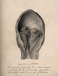
- Pictures
- Online
A human bladder with a tumour. Pencil drawing by G.E. Blenkins, 1831.
Blenkins, George Eleazar, -1894.Date: 1831Reference: 5408i
- Pictures
- Online
Nicholas Byfield, a man who had a large bladder-stone. Line engraving, 1790.
Date: 30 July 1790Reference: 209i
- Pictures
- Online
Robert Short, who had a bladder-stone removed which was eight inches in circumference. Engraving, 1820.
Date: 1820Reference: 1816i
- Pictures
- Online
Robert Short, who had a bladder-stone removed which was eight inches in circumference. Engraving.
Reference: 1815i
- Pictures
- Online
A lateral incision into the hypogastrium for removal of bladder stone. Engraving by B. Prevost after L.J. Goussier.
Goussier, Louis-Jacques, 1722-1799.Date: [1762-1772]Reference: 22850i- Pictures
Cancer of the gall bladder in a 78-year old man with acute cholecystitis: stomach specimen showing (a) encompassing yellow tumour, (b) ball shaped growth in gall bladder and (c) a cholecysto-intestinal fistula, also infecting the gall bladder. Watercolour by Barbara E. Nicholson, 1953.
Nicholson, BarbaraDate: 1953Reference: 35009iPart of: Barbara Nicholson medical illustration collection.
- Pictures
- Online
The kidney (figs 1-4), bladder and penis (fig. 5) after Nuck, Bertin and Duverney. Engraving by Benard, late 18th century.
Reference: 35105i- Pictures
William Cheselden's method of operating for bladder stone. Etching by J. Mynde, 1743, after L. Heister.
Heister, Lorenz, 1683-1758.Date: [MDCCXLIII [1743]]Reference: 2823220i- Pictures
William Cheselden's method of operating for bladder stone. Etching by F. Sesone, 1749, after L. Heister.
Heister, Lorenz, 1683-1758.Date: [1749]Reference: 2839950i
- Pictures
- Online
Germain Colot performing an operation for bladder stone. Lithograph by A. Rivoulon, 1851.
Rivoulon, Antoine, 1810-1864.Date: 1851Reference: 23338i
- Pictures
A male écorché seated, exposing his abdomen, with his bladder, rectum and anus on a ledge beside him. Woodcut by François Jollat, 1545, after Étienne de La Rivière and Charles Estienne.
Estienne, Charles, 1504-approximately 1564.Date: [1557/1575]Reference: 38851i- Pictures
Catheters for urinary obstruction, and surgical instruments for removing bladder stones. Etching by F. Sesone, 1749, after L. Heister.
Heister, Lorenz, 1683-1758.Date: [1749]Reference: 2839948i- Pictures
Catheters for urinary obstruction, and surgical instruments for removing bladder stones. Etching by J. Mynde, 1743, after L. Heister.
Heister, Lorenz, 1683-1758.Date: [MDCCXLIII [1743]]Reference: 2823192i- Pictures
A stone removed from base of bladder and prostate in a 76-year old man: detail of dumb-bell shaped urinary calculus. Watercolour by Barbara E. Nicholson, 1956.
Nicholson, BarbaraDate: 1956Reference: 35717iPart of: Barbara Nicholson medical illustration collection.
- Pictures
- Online
Left, Raw's grooved catheter; right, bladder of a male. Engraving with etching.
Reference: 47700i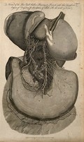
- Pictures
- Online
Nerves of the liver, gall bladder, pancreas and stomach. Line engraving (by Wooding?), 1789.
Date: Oct 17th 1789Reference: 561809i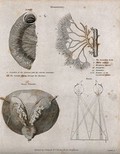
- Pictures
- Online
Mesentery: Four figures of the lacteals, the aorta & mesenterica inferior, the urinary bladder and a demonstration of vision. Line engraving by Campbell, 1816/1821.
Campbell.Date: [1816/1821]Reference: 561228i
- Pictures
- Online
Dissections of a diseased liver and gall bladder: two figures. Chromolithograph by W. Gummelt, ca. 1897.
Gummelt, W.Date: [1897?]Reference: 744178i- Pictures
Disease of female urinary bladder: scopic view. Watercolour by Barbara E. Nicholson, 1958.
Nicholson, BarbaraDate: 1958Reference: 36199iPart of: Barbara Nicholson medical illustration collection.
- Pictures
- Online
Germain Colot performing an operation for bladder stone. Coloured pencil drawing by A. Rivoulon.
Rivoulon, Antoine, 1810-1864.Reference: 22795i- Pictures
Diverticulitis in the gall bladder of a 57-year old woman with chronic cholecystitis: bladder specimen, of bilocular structure, opened to reveal stones at the fundal region. Watercolour by Barbara E. Nicholson, 1956.
Nicholson, BarbaraDate: 1956Reference: 35722iPart of: Barbara Nicholson medical illustration collection.