57 results filtered with: Digital Images

- Digital Images
- Online
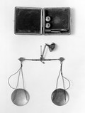
Balance and Weights, in case.
Matthias Medtmann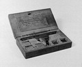
- Digital Images
- Online
Balance and Weights, in case.
Matthias Medtmann
- Digital Images
- Online
Balance and Weights, in case.
Matthias Medtmann
- Digital Images
- Online
Balance of inflammation in blood vessels, illustration
Neil Dufton
- Digital Images
- Online
Balance cases, Wellcome Chemical Research Laboratories. 19th C

- Digital Images
- Online
Balanced translocation 46,XY,t(4;10)
Wessex Reg. Genetics Centre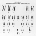
- Digital Images
- Online
Balanced translocation 45,XY,t(21;21)
Wessex Reg. Genetics Centre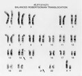
- Digital Images
- Online
Balanced translocation 45,XY,t(14;21)
Wessex Reg. Genetics Centre
- Digital Images
- Online
Balanced translocation 45,XY,t(13;14)
Wessex Reg. Genetics Centre
- Digital Images
- Online
Balanced reciprocal translocation 46,XY,t(2;5). This male has a chromosomal disorder. A chromosome 2 and a chromosome 5 have exchanged segments. The cell still contains a complete complement of
Wessex Reg. Genetics Centre
- Digital Images
- Online
Examining horse's foot balance
Royal Veterinary College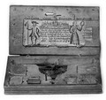
- Digital Images
- Online
Money weight balance, German. 1754
- Digital Images
- Online
Folding balance and weights, with case.

- Digital Images
- Online
Folding balance and weights, with case.

- Digital Images
- Online
Al-Jildaki, Demonstration of secrets of the balance.

- Digital Images
- Online
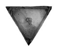
Money balance, wooden, German. circa 18th century

- Digital Images
- Online
Roman bronze balance, excavated at Pompei.

- Digital Images
- Online
Money weight balance and box weights
- Digital Images
- Online
Pan of balance for money weights showing maker's mark, label on box gives Caspar Grevenberg
- Digital Images
- Online
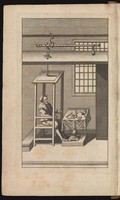
Engraving showing Sanctorio sitting in the balance that he constructed to determine the net weight change over time after the intake and excretion of food stuffs and fluids

- Digital Images
- Online
A microCT 3D reconstruction of a 10-day-old chick embryo, as seen from the right hand side. The inner ear is depicted, with the semicircular canals (the body's balance organ) and the cochlea (which converts sound waves into electrical impulses) shown in green. The otic capsule, a cartilaginous structure surrounding the inner ear which develops into part of the sphenoid bone, is shown in blue.
Akshay Kumar, Tom Davies and Nobue Itasaki, University of Bristol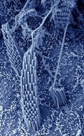
- Digital Images
- Online
Stereocilia in the vestibular organ
Dr David Furness
- Digital Images
- Online
Stereocilia in the vestibular organ
Dr David Furness
- Digital Images
- Online
Persian case of balances.

- Digital Images
- Online
Vestibular hair cells
Dr David Furness