614 results

- Archives and manuscripts
- Online
Microscope image referenced as "12 day embryonic fowl long bone after acetic"
Martin, A. V.Date: March 1952Reference: KDBP/1/1/0615Part of: King's College London Department of Biophysics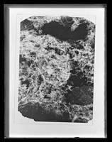
- Archives and manuscripts
- Online
Microscope image referenced as "Section of 10 day tibia "C" after hyaluronidase (1st batch spec[imen] 1)"
Martin, A. V.Date: March 1953Reference: KDBP/1/1/0919/bPart of: King's College London Department of Biophysics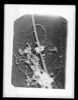
- Archives and manuscripts
- Online
Microscope image referenced as "Embryonic (18 day) RT [rat tail/tendon]. Untreated"
Martin, A. V.Date: July 1951Reference: KDBP/1/1/0388Part of: King's College London Department of Biophysics
- Archives and manuscripts
- Online
Microscope image referenced as "Fowl femur decalcified"
Martin, A. V.Date: July 1951Reference: KDBP/1/1/0413Part of: King's College London Department of Biophysics
- Archives and manuscripts
- Online
Microscope image referenced as "Pheasant leg tendon unossified intreated"
Martin, A. V.Date: July 1951Reference: KDBP/1/1/0381Part of: King's College London Department of Biophysics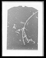
- Archives and manuscripts
- Online
Microscope image referenced as "Elephas (Siberian Mammoth) tusk decalcified. FM12"
Martin, A. V.Date: March 1952Reference: KDBP/1/1/0591Part of: King's College London Department of Biophysics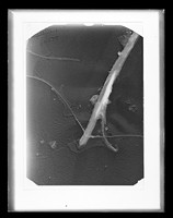
- Archives and manuscripts
- Online
Microscope image referenced as "Pheasant tendon unossified region untreated"
Martin, A. V.Date: June 1951Reference: KDBP/1/1/0342Part of: King's College London Department of Biophysics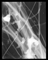
- Archives and manuscripts
- Online
Microscope image referenced as "Asterias rubens [common starfish], decalcified body wall"
Martin, A. V.Date: March 1952Reference: KDBP/1/1/0627Part of: King's College London Department of Biophysics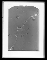
- Archives and manuscripts
- Online
Microscope image referenced as "12 day embryonic fowl long bone after acetic"
Martin, A. V.Date: March 1952Reference: KDBP/1/1/0616Part of: King's College London Department of Biophysics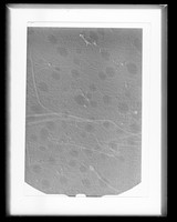
- Archives and manuscripts
- Online
Microscope image referenced as "Cartilage from 11 day chick embryo metaphysis of tibia"
Martin, A. V.Date: December 1952Reference: KDBP/1/1/0792Part of: King's College London Department of Biophysics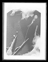
- Archives and manuscripts
- Online
Microscope image referenced as "Fowl tendon ossified region decalcified"
Martin, A. V.Date: June 1951Reference: KDBP/1/1/0345Part of: King's College London Department of Biophysics
- Archives and manuscripts
- Online
Microscope image referenced as "10 day embryonic tibia after trypsin"
Martin, A. V.Date: March 1953Reference: KDBP/1/1/0913/bPart of: King's College London Department of Biophysics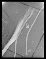
- Archives and manuscripts
- Online
Microscope image referenced as "Adult fowl leg tendon acetic acid"
Martin, A. V.Date: March 1952Reference: KDBP/1/1/0619Part of: King's College London Department of Biophysics
- Archives and manuscripts
- Online
Microscope image referenced as "Pheasant tendon unossified undecalcified"
Martin, A. V.Date: March 1951Reference: KDBP/1/1/0283Part of: King's College London Department of Biophysics
- Archives and manuscripts
- Online
Microscope image referenced as "11 day chick embryo. Under calcified frontal bone homogenate. Approx x7000"
Martin, A. V.Date: May 1950Reference: KDBP/1/1/0164Part of: King's College London Department of Biophysics
- Archives and manuscripts
- Online
Electron microscope image referenced as "Extended fibres FTU Va"
Martin, A. V.Date: July 1951Reference: KDBP/1/1/0391Part of: King's College London Department of Biophysics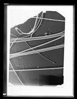
- Archives and manuscripts
- Online
Microscope image referenced as "Rat tail tendon, untreated"
Martin, A. V.Date: March 1951Reference: KDBP/1/1/0284Part of: King's College London Department of Biophysics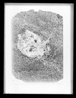
- Archives and manuscripts
- Online
Microscope image referenced as "Section of 14 day tibia head. 1st batch spec[imen] 3. Various cells and deteriorated"
Martin, A. V.Date: February 1953Reference: KDBP/1/1/0900Part of: King's College London Department of Biophysics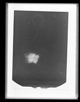
- Archives and manuscripts
- Online
Microscope image referenced as "Cytoblasts specimen"
Martin, A. V.Date: March 1952Reference: KDBP/1/1/0624Part of: King's College London Department of Biophysics
- Archives and manuscripts
- Online
Microscope image referenced as "7 day tibia head. 1st batch Specimen 1"
Martin, A. V.Date: February 1953Reference: KDBP/1/1/0817Part of: King's College London Department of Biophysics
- Archives and manuscripts
- Online
Microscope image referenced as "Young fowl neck tendon. CaCl2 collagenase"
Martin, A. V.Date: July 1951Reference: KDBP/1/1/0387Part of: King's College London Department of Biophysics
- Archives and manuscripts
- Online
Microscope image referenced as "Bacterium in division"
Martin, A. V.Date: June 1951Reference: KDBP/1/1/0340Part of: King's College London Department of Biophysics
- Archives and manuscripts
- Online
Microscope image referenced as "Fowl femur decalcified"
Martin, A. V.Date: June 1951Reference: KDBP/1/1/0341Part of: King's College London Department of Biophysics
- Archives and manuscripts
- Online
Microscope image referenced as "Young fowl neck tendon. M/1 CaCl2 collagenase"
Martin, A. V.Date: July 1951Reference: KDBP/1/1/0390Part of: King's College London Department of Biophysics
- Archives and manuscripts
- Online
Microscope image referenced as "Unidentified bacterium from decalcified preparation"
Martin, A. V.Date: March 1951Reference: KDBP/1/1/0288Part of: King's College London Department of Biophysics