2,261 results filtered with: Human anatomy

- Pictures
- Online
Nicolaus Tulp demonstrating anatomy to seven syndics of the Surgeons' Guild of Amsterdam. Etching by W. Unger after Rembrandt, 1632.
Rembrandt Harmenszoon van Rijn, 1606-1669.Reference: 544318i- Archives and manuscripts
M0007579A: Manuscript illustration of an anatomical figure from Anothomia Philippi Septimi, 1345
Date: 9 December 1940Reference: WT/D/1/20/1/65/18Part of: Wellcome Trust Corporate Archive
- Archives and manuscripts
- Online
M0007262: Male anatomical wax figure
Date: 6 September 1940Reference: WT/D/1/20/1/62/41Part of: Wellcome Trust Corporate Archive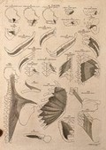
- Pictures
- Online
Muscles of the neck and trunk: 24 figures. Line engraving by A. Bell after B.S. Albinus, 1777.
Albinus, Bernhard Siegfried, 1697-1770.Date: [1777-1778]Reference: 678615i- Archives and manuscripts
M0007584A: Manuscript illustration of brain surgery from Anothomia Philippi Septimi, 1345
Date: 9 December 1940Reference: WT/D/1/20/1/65/27Part of: Wellcome Trust Corporate Archive
- Pictures
- Online
Blood vessels of the lower limb: two figures showing dissections of the leg and foot. Coloured lithograph by G.E. Madeley after A. A. Cane, 1834.
Cane, A. A.Date: [1834]Reference: 562468i
- Archives and manuscripts
- Online
M0002531: View from above and side of Ivory skeleton
Date: 10 November 1931Reference: WT/D/1/20/1/21/48Part of: Wellcome Trust Corporate Archive- Archives and manuscripts
M0007577A: Manuscript illustration of an anatomical figure from Anothomia Philippi Septimi, 1345
Date: 9 December 1940Reference: WT/D/1/20/1/65/14Part of: Wellcome Trust Corporate Archive
- Pictures
- Online
Muscleman: right lateral view. Ink drawing, 18th-19th century.
Reference: 497440i
- Pictures
- Online
Dissection of the pregnant uterus, showing the foetus at nine months. Copperplate engraving by R. Strange after I.V. Rymsdyk, 1774, reprinted 1851.
Rymsdyk, Jan van, active 1750-1788.Date: [1851]Reference: 579806iPart of: Anatomia uteri humani gravidi.- Archives and manuscripts
M0007581B: Manuscript illustration of an anatomical figure from Anothomia Philippi Septimi, 1345
Date: 9 December 1940Reference: WT/D/1/20/1/65/22Part of: Wellcome Trust Corporate Archive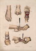
- Pictures
- Online
Six cross-sections through a foot. Coloured lithograph by M. Hanhart after C. Heath after J.B. Léveillé.
Léveillé, J. B. F. (Jean Baptiste François), 1769-1829.Date: 1875Reference: 23252i- Pictures
The spinal cord: two cross-sections. Wood engraving with ink by M. Toller (?), 1895.
Toller, M.Date: [1895]Reference: 569118i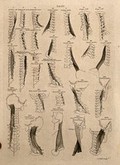
- Pictures
- Online
Muscles of the neck: 27 figures. Line engraving by A. Bell after B.S. Albinus, 1777.
Albinus, Bernhard Siegfried, 1697-1770.Date: [1777-1778]Reference: 678591i
- Pictures
- Online
The liver and the kidneys. Engraving, 1686, the third, fourth and fifth figures after G. de Lairesse, 1685.
Lairesse, Gérard de, 1640-1711.Date: [1686]Reference: 31500i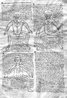
- Archives and manuscripts
- Online
M0007121: Page from an anatomical manuscript with three illustrations of the human body
Date: 30 July 1940Reference: WT/D/1/20/1/61/9Part of: Wellcome Trust Corporate Archive
- Pictures
- Online
Blood-vessels and their role in circulation of blood. Coloured lithograph by William Fairland, 1837.
Date: [1837]Reference: 641915i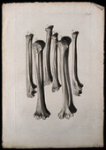
- Pictures
- Online
The bones of the arm. Engraving after G. de Lairesse, 1739.
Lairesse, Gérard de, 1640-1711.Date: 1739]Reference: 28381i
- Pictures
- Online
The arterial system: illustration of a human figure, showing the veins and arteries. Line engraving, ca. 1850.
Date: [1850?]Reference: 564545i
- Pictures
- Online
Muscles of the pelvis and leg: two écorchés, rear view. Colour mezzotint by J.F. Gautier d'Agoty after himself, 1745/1746.
Gautier Dagoty, 1717-1785.Date: [1745/1746]Reference: 572034i
- Pictures
- Online
The circulatory system: diagrams showing the heart, kidneys and pelvic bones, with the arteries and veins indicated in red and blue. Coloured lithograph by J. Maclise, 1841/1844.
Maclise, Joseph.Date: [1841/1844]Reference: 579376i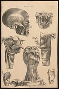
- Pictures
- Online
Muscles of the head and neck: seven figures. Line engraving, ca. 1850.
Date: [1850?]Reference: 564536i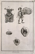
- Pictures
- Online
The uterus, a foetus, the hymen and female genitals, after Haller, Kulm and Huber. Engraving by Benard, late 18th century.
Reference: 35218i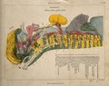
- Pictures
- Online
Torso of a woman: cross-section indicating the nerves, organs, arteries and bones, in various colours. Coloured line engraving by H. Mutlow, 1808.
Date: Sept 1st 1808Reference: 561441i
- Pictures
- Online
Dissections of the lower leg, knee joint and foot, back view: three figures, with the arteries and blood vessels indicated in red. Coloured lithograph by J. Roux, 1822.
Roux, Jacob Chr.Date: [1822]Reference: 579778iPart of: Tabulae arteriarum corporis humani.