162 results filtered with: Network

- Digital Images
- Online
Bacterial microbiome mapping, bioartistic experiment
François-Joseph Lapointe, Université de Montréal
- Digital Images
- Online
Cellular architecture of normal human skin imaged by whole mount tissue microscopy. Human skin has a rich network of white blood cells (specifically dendritic cells, T cells and macrophages) which form sheaths around blood vessels. In this image, T cells (stained for CD3; red) dendritic cells (stained for MHC class II; green) and macrophages (stained for LYVE-1; blue with some cells showing a tinge of green) can be seen. Cell nuclei have been stained with DAPI (grey). This normal cellular architecture is grossly disrupted in diseased skin (see related images). X20 magnification. Scale bar (white) represents 100 micrometres.
Dr. Xiao-nong Wang, Human Dendritic Cell Laboratory, Newcastle University
- Digital Images
- Online
Bacterial microbiome mapping, bioartistic experiment
François-Joseph Lapointe, Université de Montréal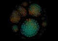
- Digital Images
- Online
Bacterial microbiome mapping, bioartistic experiment
François-Joseph Lapointe, Université de Montréal
- Digital Images
- Online
Rat neurones, SEM
Anne Weston, Francis Crick Institute
- Digital Images
- Online
Brain blood vessels, suspected meningitis, 3D printed nylon
Dave Farnham
- Digital Images
- Online
Brain blood vessels, suspected meningitis, 3D printed nylon
Dave Farnham
- Digital Images
- Online
Healthy adult human brain viewed from above, MRI
Dr Flavio Dell'Acqua
- Digital Images
- Online
Brain blood vessels, suspected meningitis, 3D printed nylon
Dave Farnham
- Digital Images
- Online
Purkinje neurons in culture
Annie Cavanagh
- Digital Images
- Online
Healthy adult human brain viewed face on, tractography
Henrietta Howells, NatBrainLab
- Digital Images
- Online
Brain blood vessels, suspected meningitis, 3D printed nylon
Dave Farnham
- Digital Images
- Online
Brain blood vessels, suspected meningitis, 3D printed nylon
Dave Farnham
- Digital Images
- Online
Brain blood vessels, suspected meningitis, 3D printed nylon
Dave Farnham
- Digital Images
- Online
Brain blood vessels, suspected meningitis, 3D printed nylon
Dave Farnham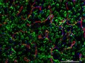
- Digital Images
- Online
Cellular architecture of normal human skin imaged by whole mount tissue microscopy. Human skin has a rich network of white blood cells (specifically dendritic cells, T cells and macrophages) which form sheaths around blood vessels. This image was taken directly beneath the junction that joins the dermal and epidermal layers of the skin (dermo-epidermal junction). At this level, the capillary network (stained for CD31; red) is visualised against a lawn of autofluorescent dermal papillae (finger-like projections of the dermis; green) scattered with dendritic cells (stained for CD11c; green) and macrophages (stained for LYVE-1; blue). This normal cellular architecture is grossly disrupted in diseased skin (see related images). Scale bar (white) represents 200 micrometres.
Dr. Xiao-nong Wang, Human Dendritic Cell Laboratory, Newcastle University
- Digital Images
- Online
Healthy adult human brain viewed from behind, tractography
Henrietta Howells, NatBrainLab
- Digital Images
- Online
Bacterial microbiome mapping, bioartistic experiment
François-Joseph Lapointe, Université de Montréal
- Digital Images
- Online
Brain blood vessels, suspected meningitis, 3D printed nylon
Dave Farnham
- Digital Images
- Online
Healthy human adult brain viewed from the side, tractography
Dr Flavio Dell'Acqua
- Digital Images
- Online
Brain blood vessels, suspected meningitis, 3D printed nylon
Dave Farnham
- Digital Images
- Online
Brain blood vessels, suspected meningitis, 3D printed nylon
Dave Farnham
- Digital Images
- Online
Cellular architecture of normal human skin imaged by whole mount tissue microscopy. Human skin has a rich network of white blood cells (specifically dendritic cells, T cells and macrophages) which form sheaths around blood vessels. In this image, blood vessels (string-like structures stained for CD31; green), lymphatic vessels (ribbon-like structures stained for LYVE-1; blue) and T cells (stained for CD3; red) can be seen. T cells are only found around dermal blood vessels. Macrophages (stained for LYVE-1; blue) are also present. This normal cellular architecture is grossly disrupted in diseased skin (see related images). X10 magnification. Scale bar (white) represents 200 micrometres.
Dr. Xiao-nong Wang, Human Dendritic Cell Laboratory, Newcastle University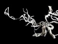
- Digital Images
- Online
Brain blood vessels, suspected meningitis, 3D printed nylon
Dave Farnham
- Digital Images
- Online
Brain blood vessels, suspected meningitis, 3D printed nylon
Dave Farnham