323 results
- Pictures
Stomach cancer in a 58-year old man with gastric ulcer: (a) view of stomach from resected upper end and (b) view of opened specimen, from incision along greater curvature. Watercolour by Barbara E. Nicholson, 1950.
Nicholson, BarbaraDate: 1950Reference: 33895iPart of: Barbara Nicholson medical illustration collection.- Pictures
Bacterial endocarditis of mitral valve and aorta in a 62-year old man with congestive heart failure: sections of heart to show (a) vegetation on aortic valve and (b) ulcerated aortic cusps. Watercolour by Barbara E. Nicholson, 1952.
Nicholson, BarbaraDate: 1952Reference: 34500iPart of: Barbara Nicholson medical illustration collection.- Pictures
Dissecting aneurysm of aorta in a male patient: section showing (a) aortic valve and extensively diseased descending aorta cloaked in large blood clot, with (b) inset of independant thrombus in carotid artery. Watercolour by Barbara E. Nicholson, 1958.
Nicholson, BarbaraDate: 1958Reference: 36031iPart of: Barbara Nicholson medical illustration collection.- Pictures
Fatty replacement of kidney in a 52-year old man: sections showing (a) adipose tissue, which has entirely consumed the kidney and (b) small cavity, representing renal pelvis, which contained one calculus. Watercolour by Barbara E. Nicholson, 1958.
Nicholson, BarbaraDate: 1958Reference: 36029iPart of: Barbara Nicholson medical illustration collection.- Pictures
Aneurysm of splenic artery in an 82-year old woman with pneumonia and fatal congestive heart failure: specimen showing (a) two unruptured but grossly calcified aneurysms with (b) tortuous atheromatous splenic artery. Watercolour by Barbara E. Nicholson, 1958.
Nicholson, BarbaraDate: 1958Reference: 36034iPart of: Barbara Nicholson medical illustration collection.- Pictures
Aneurysm of abdominal aorta in a 73-year old man with fatal hypertensive heart failure: (a) diagram of abdomen, to scale after radiograph and (b) sketch showing calcified masses. Pen and ink drawing by Barbara E. Nicholson, 1953.
Nicholson, BarbaraDate: 1953Reference: 34855iPart of: Barbara Nicholson medical illustration collection.- Pictures
Mitral valve disease in a 63-year old man with congestive heart failure: detail of (a) left atrium and (b) left ventricle showing enlargement disease in the edges of the anterior cusp. Watercolour by Barbara E. Nicholson, 1952.
Nicholson, BarbaraDate: 1952Reference: 34438iPart of: Barbara Nicholson medical illustration collection.- Pictures
Obstructed ileum in a male patient with fatal pulmonary embolism: surgical specimen showing (a) localised narrowing and thickening of the bowel wall, and (b) granulation tissue replacing mucuosa and the muscular coat. Watercolour by Barbara E. Nicholson, 1951.
Nicholson, BarbaraDate: 1951Reference: 34312iPart of: Barbara Nicholson medical illustration collection.- Books
The Royal Albert Institution, Lancaster : for the feeble-minded belonging to Lancashire, Yorkshire, Cheshire, Westmorland, Cumberland, Durham and Northumberland fifty-first annual report.
Royal Albert Institution (Lancaster, England)Date: 1915- Pictures
Fatal haemopericardium in a 70-year old woman with dyspnoea and posterior infarction: specimen showing (a) rupture, from base of posterior papillary muscle internally across the pericardium surface, and (b) recent thrombosis in right coronary. Watercolour by Barbara E. Nicholson, 1954.
Nicholson, BarbaraDate: 1954Reference: 35226iPart of: Barbara Nicholson medical illustration collection.- Pictures
Schonlein's purpura in a 50-year old woman: sketch of (a) legs, anterior view and (b) tongue, showing swollen joints and discolouration (resulting from extravasation of blood into the skin and mucus membranes). Watercolour by Barbara E. Nicholson, 1951.
Nicholson, BarbaraDate: 1951Reference: 34396iPart of: Barbara Nicholson medical illustration collection.- Pictures
Fatal haemopericardium in a 73-year old woman with coronary disease and diabetes: heart and section showing (a) ruptured ventricle which caused death and (b) thrombosis of anterior descending branch of left coronary. Watercolour by Barbara E. Nicholson, 1954.
Nicholson, BarbaraDate: 1954Reference: 35183iPart of: Barbara Nicholson medical illustration collection.- Books
The Royal Albert Institution, Lancaster : for the feeble-minded belonging to Lancashire, Yorkshire, Cheshire, Westmorland, Cumberland, Durham and Northumberland fiftieth annual report.
Royal Albert Institution (Lancaster, England)Date: 1914- Pictures
Fatal adrenal pheochromocytoma in a 48-year woman with hypertension and pontile haemorrhage: (a) section of left kidney, displaced by chromaffin tumour with gross exudates and (b) tracing after intravenous pyelogram showing pelvic pattern. Watercolour by Barbara E. Nicholson, 1954.
Nicholson, BarbaraDate: 1954Reference: 35313iPart of: Barbara Nicholson medical illustration collection.- Pictures
Widespread cancer in a 50-year old man with fatal bronchopneumonia: detail sections of (a) left kidney, with fungus like growths, (b) left heart showing metastases nodules, and (c) right heart encompassed in yellow tumours. Watercolour by Barbara E. Nicholson, 1951.
Nicholson, BarbaraDate: 1951Reference: 34318iPart of: Barbara Nicholson medical illustration collection.- Pictures
Intestinal cancer and mesenteric thrombosis in a 68-year old man with infarcted gut and gangrene: diagram demonstrating conduction of blood in relation to (a) venous obstruction and (b) tumorous occlusion. Pen and ink sketch by Barbara E. Nicholson, 1957.
Nicholson, BarbaraDate: 1957Reference: 35827iPart of: Barbara Nicholson medical illustration collection.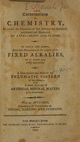
- Books
- Online
Conversations on chemistry : in which the elements of that science are familiarly explained and illustrated by experiments and plates : to which are added, some late discoveries on the subject of the fixed alkalies / by H. Davy ... ; a description and plate of the pneumatic cistern of Yale College ; and, a short account of artificial mineral waters in the United States ; with an appendix, consisting of treatises on dyeing, tanning, and currying.
Marcet, Mrs. (Jane Haldimand), 1769-1858.Date: 1809- Books
The Royal Albert Institution, Lancaster : for the feeble-minded belonging to Lancashire, Yorkshire, Cheshire, Westmorland, Cumberland, Durham and Northumberland forty-seventh annual report.
Royal Albert Institution (Lancaster, England)Date: 1911- Pictures
Congestive heart failure in a 70-year old man with digitalis, dyspnoea and chronic abdominal distension: surgical specimens of (a) embolism in superior mesenteric artery, (b) intra-mural thrombus in left ventricle and (c) diagram of gangrenous small intestine. Watercolour by Barbara E. Nicholson, 1951.
Nicholson, BarbaraDate: 1951Reference: 34211iPart of: Barbara Nicholson medical illustration collection.
- Books
- Online
The Royal Albert Institution, Lancaster : for the feeble-minded belonging to Lancashire, Yorkshire, Cheshire, Westmorland, Cumberland, Durham and Northumberland forty-sixth annual report.
Royal Albert Institution (Lancaster, England)Date: 1910- Pictures
Calcified aortic stenosis in an 80-year old woman with diabetes and fatal pulmonary oedema: section of mitral valve and aorta showing (a) rigid calcified cusps and (b) gross narrowing of the coronary vessels due to calcification. Watercolour by Barbara E. Nicholson, 1954.
Nicholson, BarbaraDate: 1954Reference: 35172iPart of: Barbara Nicholson medical illustration collection.- Pictures
Malignant abdominal tumour in a 65-year old woman with dyspepsia and haematemesis: sections of (a) spleen, opened to show large egg-shaped sarcoma and (b) diaphragm detail showing pendulous tumour attachments, the primary site of cancer. Watercolour by Barbara E. Nicholson, 1958.
Nicholson, BarbaraDate: 1958Reference: 36027iPart of: Barbara Nicholson medical illustration collection.- Pictures
Dissecting aortic aneurysm in a 60-year old man with fatal posterior cardiac infarction: (a) section of collapsed heart and (b) pen and ink sketch of abdominal aorta showing large clot, burst into the posterior mediastinium. Watercolour by Barbara E. Nicholson, 1955.
Nicholson, BarbaraDate: 1955Reference: 35420iPart of: Barbara Nicholson medical illustration collection.- Pictures
Rodent ulcer in an 82-year old man with cancer: (a) detail sketch of ulcer on left tagus and flat scabby ulceration above external auditory meatus with (b) inset to show ulceration behind ear, pinna pulled forward. Watercolour by Barbara E. Nicholson, 1957.
Nicholson, BarbaraDate: 1957Reference: 35829iPart of: Barbara Nicholson medical illustration collection.- Pictures
Schoenlein's purpura in a 50-year old woman: sketch of (a) buttocks and legs and (b) hands, anterior and posterior views, showing swollen joints and discolouration (resulting from extravasation of blood into the skin and mucus membranes). Watercolour by Barbara E. Nicholson, 1951.
Nicholson, BarbaraDate: 1951Reference: 34320iPart of: Barbara Nicholson medical illustration collection.