1,008 results filtered with: Pictures, Digital Images

- Pictures
- Online
Dissections of the male urogenital system and pelvic region: four figures, with the arteries and blood vessels indicated in red. Coloured lithograph by J. Roux, 1822.
Roux, Jacob Chr.Date: [1822]Reference: 579772iPart of: Tiedemann, Friedrich, 1781-1861.
- Pictures
- Online
Dissections of a mole: nine figures, showing the skeleton and musculature of the underside of the animal. Lithograph by R. Mintern after F.W. Brookman, 1880/1900?.
Brookman, F. W.Date: [1880/1900?]Reference: 571449i
- Pictures
- Online
Dissections of the lower leg, knee joint and foot, back view: three figures, with the arteries and blood vessels indicated in red. Coloured lithograph by J. Roux, 1822.
Roux, Jacob Chr.Date: [1822]Reference: 579778iPart of: Tiedemann, Friedrich, 1781-1861.
- Pictures
- Online
Dissections of abortions of around eight and nine weeks: six figures, each with an accompanying line diagram. Copperplate engraving by T. Worlidge after J.V. Rymsdyk, 1774, reprinted 1851.
Rymsdyk, Jan van, active 1750-1788.Date: [1851]Reference: 579882iPart of: Hunter, William
- Pictures
- Online
Dissections of the shoulder and arm; two figures, one showing a tumour or growth (?), with the arteries and blood vessels indicated in red. Coloured lithograph by J. Roux, 1822.
Roux, Jacob Chr.Date: [1822]Reference: 579755iPart of: Tiedemann, Friedrich, 1781-1861.
- Pictures
- Online
Dissections of the pregnant uterus and the corpus luteum in the left overy, at five months: three figures. Copperplate engraving by E. Du Mesnil after J.V. Rymsdyk, 1774, reprinted 1851.
Rymsdyk, Jan van, active 1750-1788.Date: [1851]Reference: 579879iPart of: Hunter, William
- Pictures
- Online
Dissections of the pregnant uterus at three months, showing the foetus in the uterus and a section of the placenta and uterus, with the foetus attached by the umbilical cord: three figures, including an outline diagram. Copperplate engraving by J.V. Rymsdyk after himself, 1774, reprinted 1851.
Rymsdyk, Jan van, active 1750-1788.Date: [1851]Reference: 579880iPart of: Hunter, William- Digital Images
Deep View of the dissection of the Pterygoid Region.

- Pictures
- Online
Face and brain: dissections. Colour mezzotint by J.F. Gautier d'Agoty, 1748.
Gautier Dagoty, 1717-1785.Date: [1748]Reference: 560776i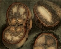
- Pictures
- Online
The brain: three dissections. Colour mezzotint by J.F. Gautier d'Agoty, 1748.
Gautier Dagoty, 1717-1785.Date: [1748]Reference: 560758i
- Pictures
- Online
Brain of a ferret: dissections with accompanying notes. Watercolour, possibly by D. Gascoigne Lillie, ca. 1905.
Lillie, Denis Gascoigne, 1888-1963.Date: 1905Reference: 572972i
- Pictures
- Online
The heart: three dissections. Colour mezzotint by J. F. Gautier d'Agoty after himself, 1754.
Gautier Dagoty, 1717-1785.Date: [1754]Reference: 572092i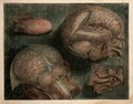
- Pictures
- Online
Tongue, brain, nasal cavity: two dissections. Colour mezzotint by J.F. Gautier d'Agoty, 1748.
Gautier Dagoty, 1717-1785.Date: [1748]Reference: 560771i
- Pictures
- Online
The human eye: fifteen figures, showing dissections of the eye. Engraving by T. Milton, 1811.
Date: Feb. 1st 1811Reference: 583172iPart of: Human and animal anatomy: album containing a collection of engravings.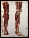
- Pictures
- Online
Two dissections showing the muscles of the leg. Watercolour by J.C. Zeller, ca. 1833.
Zeller, Johann Conrad, 1807-1856.Date: [1833?]Reference: 581467i- Pictures
Three anatomical dissections taking place in an attic. Coloured lithograph by T. C. Wilson after a pen and wash drawing by T. Rowlandson.
Rowlandson, Thomas, 1756-1827.Reference: 25405i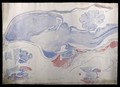
- Pictures
- Online
Brain of a chick: four figures showing dissections of the brain. Watercolour, possibly by D. Gascoigne Lillie, ca. 1905.
Lillie, Denis Gascoigne, 1888-1963.Date: 1905Reference: 572974i
- Pictures
- Online
One fascicule of life-sized lithographs, showing dissections carried out by George Viner Ellis: front of wrapper. Letterpress, 1867.
Ford, G. H. (George Henry)Date: [1867]Reference: 568655i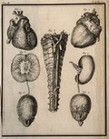
- Pictures
- Online
Wax models of the viscera, etc. by Jean-Joseph Sue père, after his own dissections. Engraving, 1749.
Date: [1749]Reference: 34226i
- Pictures
- Online
Sexual organs, male and female: ten figures of dissections. Line engraving by Lodge after F. Birnie, 1789.
Birnie, Frederick.Date: Aug 28th 1789Reference: 561800i
- Pictures
- Online
The circulatory system: dissections of the aortic arch (?), thirteen figures. Coloured lithograph by J. Maclise, 1841/1844.
Maclise, Joseph.Date: [1841/1844]Reference: 579505i
- Pictures
- Online
Brain of an eagle: figures showing dissections of the brain. Watercolour and ink, possibly by D. Gascoigne Lillie, ca 1906.
Lillie, Denis Gascoigne, 1888-1963.Date: 1906Reference: 573012i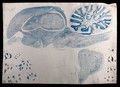
- Pictures
- Online
Brain of a guillemot: figures showing dissections and microscopic views of the brain. Watercolour, possibly by D. Gascoigne Lillie, ca 1906.
Lillie, Denis Gascoigne, 1888-1963.Date: 1906Reference: 573014i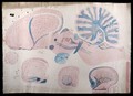
- Pictures
- Online
Brain of a rhoea (ie. rhea): five figures showing dissections of the brain. Watercolour, possibly by D. Gascoigne Lillie, ca 1906.
Lillie, Denis Gascoigne, 1888-1963.Date: 1906Reference: 573011i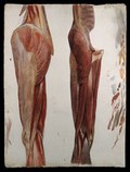
- Pictures
- Online
Two dissections showing the muscles of the thigh, hip, pelvis and knee. Watercolour by J.C. Zeller, ca. 1833.
Zeller, Johann Conrad, 1807-1856.Date: [1833?]Reference: 581459i