737 results

- Digital Images
- Online
Small blood vessel - endothelial cells & RBC
Rob Young- Ephemera
Life blood : Mr. Peter Emblem (31), whose story appears inside, suffered from a condition in which the bone marrow stops producing blood cells.
Date: 1956
- Digital Images
- Online
Cellular architecture of normal human skin imaged by whole mount tissue microscopy. Human skin has a rich network of white blood cells (specifically dendritic cells, T cells and macrophages) which form sheaths around blood vessels. This image was taken greater than 150 micrometres beneath the junction that joins the dermal and epidermal layers of the skin (dermo-epidermal junction). At this level, dendritic cells (stained for CD11c; green) and macrophages (stained for LYVE-1; blue) form clusters around blood vessels (stained for CD31; red). This normal cellular architecture is grossly disrupted in diseased skin (see related images). Scale bar (white) represents 100 micrometres.
Dr. Xiao-nong Wang, Human Dendritic Cell Laboratory, Newcastle University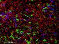
- Digital Images
- Online
Cellular architecture of human skin lymphoma imaged by whole mount tissue microscopy. Normal human skin has a rich network of white blood cells (specifically dendritic cells, T cells and macrophages) which form sheaths around blood vessels. In diseased skin, such as in skin lymphoma as seen here, this normal architecture becomes distorted. In this image, lots of T cells (stained for CD3; red), dendritic cells (stained for CD11c; green) and macrophages (stained for LYVE-1; blue) have infiltrated the skin. X20 magnification. Scale bar (white) represents 100 micrometres.
Dr. Xiao-nong Wang, Human Dendritic Cell Laboratory, Newcastle University
- Digital Images
- Online
Cellular architecture of normal human skin imaged by whole mount tissue microscopy. Human skin has a rich network of white blood cells (specifically dendritic cells, T cells and macrophages) which form sheaths around blood vessels. This image was taken less than 20 micrometres beneath the junction that joins the dermal and epidermal layers of the skin (dermo-epidermal junction). At this level, dendritic cells (stained for CD11c; green) form clusters around and between blood capillary loops (stained for CD31; red). The blind-ended tips of initial lymphatic vessels are just visible (stained for LYVE-1; blue) at this level. This normal cellular architecture is grossly disrupted in diseased skin (see related images). Scale bar (white) represents 200 micrometres.
Dr. Xiao-nong Wang, Human Dendritic Cell Laboratory, Newcastle University
- Digital Images
- Online
Choroidal vessels in the human eye with red blood cells
Peter Maloca- Books
Ultrastructure of haemic cells : a cytological atlas of normal and leukaemic blood and bone marrow / J.C. Cawley, F.G.J. Hayhoe.
Cawley, J. C.Date: 1973
- Books
- Online
On the existence, within the liver cells, of canaliculi which are in direct communication with the blood capillaries / by E.A. Schäfer.
Sharpey-Schäfer, E. A. (Edward Albert), Sir, 1850-1935.Date: [1902]- Archives and manuscripts
Oxford: technical blood grouping items
Date: mid-late 20th centuryReference: SA/HHC/R/2Part of: Harrison-Howell Blood Transfusion Collection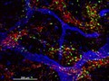
- Digital Images
- Online
Cellular architecture of normal human skin imaged by whole mount tissue microscopy. Human skin has a rich network of white blood cells (specifically dendritic cells, T cells and macrophages) which form sheaths around blood vessels (string-like structures). A network of lymphatic vessels (ribbon-like structures) is also present. In this image, human skin lymphatic vessels (stained for LYVE-1; blue) and white blood cells comprised of dendritic cells (stained for CD11c; green) and T cells (stained for CD3; red) can be seen. Some macrophages also express the protein LYVE-1 similar to lymphatic vessel cells which can be appreciated as blue cells within and in between the sheaths of white blood cells. This normal cellular architecture is grossly disrupted in diseased skin (see related images). X10 magnification. Scale bar (white) represents 200 micrometres.
Dr. Xiao-nong Wang, Human Dendritic Cell Laboratory, Newcastle University- Pictures
Shigellosis: acute: microscopic view of macrophage cells (white blood cells). Drawing by P.H. Manson-Bahr, ca. 1930.
Manson-Bahr, Philip H. (Philip Henry), Sir, 1881-1966.Date: 1930Reference: 571049i- Books
The biology of the blood-cells : with a glossary of hæmatological terms ; for the use of practitioners of medicine / by O.C. Gruner.
Gruner, O. Cameron (Oskar Cameron), 1877-1972.Date: 1913
- Digital Images
- Online
Avian blood
Royal Veterinary College
- Digital Images
- Online
Avian blood
Royal Veterinary College
- Digital Images
- Online
Avian blood
Royal Veterinary College
- Digital Images
- Online
Avian blood
Royal Veterinary College- Books
Transport and diffusion in red blood cells : by R. Whittam.
Whittam, R.Date: 1964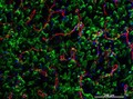
- Digital Images
- Online
Cellular architecture of normal human skin imaged by whole mount tissue microscopy. Human skin has a rich network of white blood cells (specifically dendritic cells, T cells and macrophages) which form sheaths around blood vessels. This image was taken directly beneath the junction that joins the dermal and epidermal layers of the skin (dermo-epidermal junction). At this level, the capillary network (stained for CD31; red) is visualised against a lawn of autofluorescent dermal papillae (finger-like projections of the dermis; green) scattered with dendritic cells (stained for CD11c; green) and macrophages (stained for LYVE-1; blue). This normal cellular architecture is grossly disrupted in diseased skin (see related images). Scale bar (white) represents 200 micrometres.
Dr. Xiao-nong Wang, Human Dendritic Cell Laboratory, Newcastle University
- Digital Images
- Online
Avian blood
Royal Veterinary College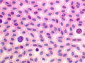
- Digital Images
- Online
Avian blood
Royal Veterinary College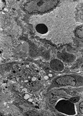
- Digital Images
- Online
Cell death around scleral blood vessels
Rob Young- Pictures
Six examples of blood cells, showing histological elements of normal blood, with additional samples displaying symptoms of various medical conditions, as seen through a microscope. Colour lithograph by R. Muir, ca. 1906.
Muir, Richard (Bacteriologist)Date: [1906?]Reference: 576153i- Pictures
Blood capillaries (?) and cells: three microscopical images illustrating the anatomy of the blood, including an illustration of the capillaries (?) . Colour lithograph by F. Reichhold, 1890/1910?.
Date: [1890/1910?]Reference: 569245i- Books
Selective action of x-rays on the blood cells of the cat / by Samson Wright and H.A. Bulman.
Wright, Samson.Date: [1929]
- Digital Images
- Online
Blood vessels in the retina showing the endothelial cells in red and the vascular contents in green. Surrounding cell nuclei are stained blue.
Freya Mowat