363 results filtered with: Pictures

- Pictures
- Online
Vessels and muscles of the arm and hand: two figures showing dissections of a left and right arm and hand, with palms facing upwards. Coloured lithograph by G.E. Madeley after A. A. Cane, 1834.
Cane, A. A.Date: [1834]Reference: 562436i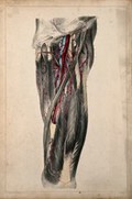
- Pictures
- Online
Blood vessels of the thigh. Coloured lithograph by G.E. Madeley after A. A. Cane, 1834.
Cane, A. A.Date: [1834]Reference: 562461i
- Pictures
- Online
Blood-vessels of the human body: nine figures. Line engraving by I. Taylor, 1790/1810(?).
Date: [1790?/1810?]Reference: 678510i
- Pictures
- Online
Surgery of blood vessels of the lower limb: four figures showing incisions in the leg and foot, with surgical instruments indicating blood vessels. Coloured lithograph by G.E. Madeley after A. A. Cane, 1834.
Cane, A. A.Date: [1834]Reference: 562472i
- Pictures
- Online
The arteries and lungs (fig. 2), after Haller; the breast (fig. 3), after Nuck; branch of the bronchi (fig. 4), after Bidloo. Engraving by Prevost, 1762.
Date: [1762]Reference: 36253i
- Pictures
- Online
The arteries and lungs (fig. 2), after Haller; the breast (fig. 3), after Nuck; branch of the bronchi (fig. 4), after Bidloo. Engraving by Benard, late 18th century.
Reference: 35078i
- Pictures
- Online
The arteries of the head after Haller; the eye, after Ruysch; the tongue after Heister. Engraving by Prevost, 1762.
Rollinus, C. J., active 18th century.Date: [1762]Reference: 35484i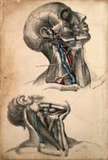
- Pictures
- Online
Muscles and blood vessels of the head and neck: two figures of a dissection. Coloured lithograph by G.E. Madeley after A.A. Cane, 1834.
Cane, A. A.Date: [1834]Reference: 562428i
- Pictures
- Online
The arteries of the head after Haller; the eye, after Ruysch; the tongue after Heister. Engraving by Benard, late 18th century.
Rollinus, C. J., active 18th century.Reference: 34490i
- Pictures
- Online
Anatomy and botany; top left, dissected head and chest showing arteries; top right, larynx; bottom left, buckthorn plant; centre, surgical instruments; bottom right, electric ray fish. Coloured engraving, 1834-1837.
Date: [between 1834 and 1837]Reference: 577936i
- Pictures
- Online
Anatomy and botany: top left, arteries in thorax and abdomen; top right, superior section of the brain; centre left, lateral distortion owing to chronic pleurisy; centre right, part of the lung; bottom, foxglove and aconite. Coloured engraving, 1834-1837.
Date: [between 1834 and 1837]Reference: 577933i
- Pictures
- Online
Subclavian and axillary vessels: dissection. Coloured lithograph by G.E. Madeley after A. A. Cane, 1834.
Cane, A. A.Date: [1834]Reference: 562431i
- Pictures
- Online
The arteries of the head after Haller; the eye, after Ruysch, Cowper and Bidloo. Engraving by A.J. Defehrt, 1762.
Rollinus, C. J., active 18th century.Date: [1762]Reference: 35483i
- Pictures
- Online
Arteries, veins and bones: fourteen figures. Line engraving by J. Record, 1780/1790?.
Date: [1780/1790?]Reference: 561604i
- Pictures
- Online
The arteries of the head after Haller; the eye, after Ruysch, Cowper and Bidloo. Engraving by Benard, late 18th century.
Rollinus, C. J., active 18th century.Reference: 34482i
- Pictures
- Online
Anatomy, surgery and botany; top, dissected head showing arteries; centre, methods of bandaging the thorax and head; below, cleft lip (harelip) and instruments for surgery on it; bottom, fly agaric mushroom. Coloured engraving, 1834-1837.
Date: [between 1834 and 1837]Reference: 577937i
- Pictures
- Online
Ambroise Paré using the ligature when amputating at the siege of Damvillers, 1552. Oil painting by Ernest Board.
Board, Ernest, 1877-1934.Reference: 45900i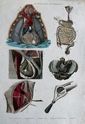
- Pictures
- Online
Top left, section of the heart; top right, intestines; centre left and right, hernia; bottom left, carotide; bottom right, speculum. Coloured engraving, 1834-1837.
Date: [between 1834 and 1837]Reference: 577932i
- Pictures
- Online
The arterial system: illustration of a human figure, showing the veins and arteries. Line engraving, ca. 1850.
Date: [1850?]Reference: 564545i- Pictures
Aortic aneurysm in a 63-year old man with oedema, colick and ecchymiosis in left loin: section of abdominal aorta, just above level of renal arteries, showing two to three centimetres of laminated clot. Watercolour by Barbara E. Nicholson, 1952.
Nicholson, BarbaraDate: 1952Reference: 34446iPart of: Barbara Nicholson medical illustration collection.- Pictures
Aortic aneurysm in a 63-year old man with oedema, colick and ecchymiosis in left loin: specimen and section of abdominal aorta, just above level of renal arteries, showing two to three centimetres of laminated clot. Watercolour by Barbara E. Nicholson, 1952.
Nicholson, BarbaraDate: 1952Reference: 34439iPart of: Barbara Nicholson medical illustration collection.- Pictures
Atheroma in a 68-year old man with perforated stomach ulcer and fatal uraemia: section showing gross deterioration of arteries with dissecting aneurysm. Watercolour by Barbara E. Nicholson, 1950.
Nicholson, BarbaraDate: 1950Reference: 33548iPart of: Barbara Nicholson medical illustration collection.
- Pictures
- Online
The arterial system of the human body. Engraving, 1568.
Becerra, Gaspar, 1520?-1568?Reference: 26965i
- Pictures
- Online
The pulmonary veins and arteries. Engraving, 1686.
Date: [1686]Reference: 29930i
- Pictures
- Online
The course of the veins and the arteries through the body (Table IV), after Eustachius; the arterial system (Table V), after Cowper in Drake. Etching by I. Basire, 1743.
Date: [1743]Reference: 37028i