1,008 results filtered with: Pictures, Digital Images

- Pictures
- Online
Dissections of abortions of around eight and nine weeks: six figures, each with an accompanying line diagram. Copperplate engraving by T. Worlidge after J.V. Rymsdyk, 1774, reprinted 1851.
Rymsdyk, Jan van, active 1750-1788.Date: [1851]Reference: 579882iPart of: Anatomia uteri humani gravidi.
- Pictures
- Online
Dissections of two diseased hearts. Chromolithograph by W. Gummelt, ca. 1897.
Gummelt, W.Date: [1897?]Reference: 577221i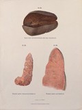
- Pictures
- Online
Dissections of diseased spleen: three figures. Chromolithograph by W. Gummelt, ca. 1897.
Gummelt, W.Date: [1897?]Reference: 577065i
- Pictures
- Online
Dissections of diseased lungs affected by emphysema. Chromolithograph by W. Gummelt, ca. 1897.
Gummelt, W.Date: [1897?]Reference: 577580i
- Pictures
- Online
Dissections of pharynxes and larynxes affected by diseases including syphilis. Chromolithograph by W. Gummelt, ca. 1897.
Gummelt, W.Date: [1897?]Reference: 577578i
- Pictures
- Online
Dissections of the male genital and rectal area: two figures. Coloured wood engraving with letterpress, 1860/1900?.
Date: [1860?/1900?]Reference: 568836i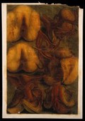
- Pictures
- Online
Dissections of the female urogenital system: six figures. Colour mezzotint by J. F. Gautier d'Agoty after himself, 1754.
Gautier Dagoty, 1717-1785.Date: [1754]Reference: 572095i
- Pictures
- Online
Dissections of chicks of various species: eleven figures, including details of the feet. Lithograph by J. Erxleben (?), 1840/1860?.
Erxleben, James, active 1839-1852.Date: [1840/1860?]Reference: 571033i
- Pictures
- Online
Dissections of an elephant's foot and tail: seven figures. Etching by R. Vinkeles 1787/1800 (?), after P. Camper, 1774.
Camper, Petrus, 1722-1789.Date: [1787/1800?]Reference: 571094i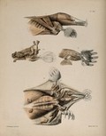
- Pictures
- Online
Dissections of a mole: four figures, showing the muscles of the head and limbs. Lithograph by F.W. Brookman, 1880/1900?.
Brookman, F. W.Date: [1880/1900?]Reference: 571450i
- Pictures
- Online
Dissections of the stomach: two figures, with the arteries and blood vessels indicated in red. Coloured lithograph by J. Roux, 1822.
Roux, Jacob Chr.Date: [1822]Reference: 579764iPart of: Tabulae arteriarum corporis humani.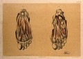
- Pictures
- Online
Dissections of the underside of the foot, showing the muscles and blood vessels: two figures. Colour lithograph by G.H. Ford, 1867.
Ford, G. H. (George Henry)Date: 1867Reference: 580546i
- Pictures
- Online
Dissections of femoral arteries affected by calcification, with illustrations of subperitoneal phlebectasia and rectal rectal hemorrhoids. Chromolithograph by W. Gummelt, ca. 1897.
Gummelt, W.Date: [1897?]Reference: 577242i
- Pictures
- Online
Dissections of the arm and hand; four figures, with the arteries and blood vessels indicated in red. Coloured lithograph by J. Roux, 1822.
Roux, Jakob Wilhelm Christian, 1775-1831.Date: [1822]Reference: 579759iPart of: Tabulae arteriarum corporis humani.
- Pictures
- Online
Dissections of the arm, hand and elbow joint; three figures, with the blood vessels indicated in red. Coloured lithograph by J. Roux, 1822.
Roux, Jakob Wilhelm Christian, 1775-1831.Date: [1822]Reference: 579753iPart of: Tabulae arteriarum corporis humani.
- Pictures
- Online
Dissections of the arm and elbow; three figures, with the arteries and blood vessels indicated in red. Coloured lithograph by J. Roux, 1822.
Roux, Jakob Wilhelm Christian, 1775-1831.Date: [1822]Reference: 579757iPart of: Tabulae arteriarum corporis humani.
- Pictures
- Online
Dissections of the pregnant uterus at five months: two figures. Copperplate engraving by P.C. Canot after J.V. Rymsdyk, 1774, reprinted 1851.
Rymsdyk, Jan van, active 1750-1788.Date: [1851]Reference: 579836iPart of: Anatomia uteri humani gravidi.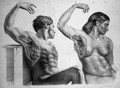
- Pictures
Dissections of the muscles and blood-vessels of the axilla of a seated man and a seated woman. Coloured lithograph by J. Maclise, 1851.
Maclise, Joseph.Date: [1851]Reference: 640716i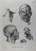
- Pictures
- Online
Dissections of the face, neck and jaw: five figures, with the arteries and blood vessels indicated in red. Coloured lithograph by J. Roux, 1822.
Roux, Jakob Wilhelm Christian, 1775-1831.Date: [1822]Reference: 579725iPart of: Tabulae arteriarum corporis humani.
- Pictures
- Online
Dissections of the male genitalia and upper thighs: four figures, with the arteries and blood vessels indicated in red. Coloured lithograph by J. Roux, 1822.
Roux, Jacob Chr.Date: [1822]Reference: 579776iPart of: Tabulae arteriarum corporis humani.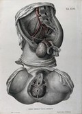
- Pictures
- Online
Dissections of the female anus and urogenital system: two figures, with the arteries and blood vessels indicated in red. Coloured lithograph by J. Roux, 1822.
Roux, Jakob Wilhelm Christian, 1775-1831.Date: [1822]Reference: 579768iPart of: Tabulae arteriarum corporis humani.
- Pictures
- Online
Dissections of a mole: thirteen figures, including the musculature of the head, neck, legs and feet. Lithograph by R. Mintern after F.W. Brookman, 1880/1900?.
Brookman, F. W.Date: [1880/1900?]Reference: 571437i
- Pictures
- Online
Dissections of the male urogenital system and pelvic region: four figures, with the arteries and blood vessels indicated in red. Coloured lithograph by J. Roux, 1822.
Roux, Jacob Chr.Date: [1822]Reference: 579772iPart of: Tabulae arteriarum corporis humani.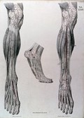
- Pictures
- Online
Dissections of the lower leg and the foot, front view: three figures, with the arteries and blood vessels indicated in red. Coloured lithograph by J. Roux, 1822.
Roux, Jacob Chr.Date: [1822]Reference: 579777iPart of: Tabulae arteriarum corporis humani.
- Pictures
- Online
Dissections of the head of a mole: three figures, showing the musculature of the animal's head and neck. Lithograph by E. Wilson and F.W. Brookman, 1880/1900?.
Brookman, F. W.Date: [1880/1900?]Reference: 571433i