2,381 results
- Books
Dissection : photographs of a rite of passage in American medicine, 1880-1930 / John Harley Warner, James M. Edmonson.
Warner, John Harley, 1953-Date: 2009
- Pictures
- Online
Dissection of a horse's lungs and heart: three figures. Coloured engraving by J. Pass after Harguinier, 1805.
Harguinier, active 1763-1768.Date: 1805Reference: 570690i
- Pictures
- Online
Dissection of a horse's foot. Engraving with etching and stipple by W. Skelton after St. Aubin, 1800/1830?.
St. Aubin.Date: [1800/1830?]Reference: 570754i- Pictures
Dissection of the abdomen, showing the viscera, blood vessels and arteries. Lithograph by P. Highley, 1907.
Highley, Percy.Date: [1907]Reference: 568757i- Pictures
Dissection of the abdomen, showing the viscera, blood vessels and arteries. Lithograph by P. Highley, 1907.
Highley, Percy.Date: [1907]Reference: 568758i
- Pictures
- Online
Dissection of muscles and blood-vessels of the upper arm: two figures. Coloured lithograph by J. Maclise, 1851.
Maclise, Joseph.Date: [1851]Reference: 640717i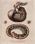
- Pictures
- Online
Dissection of a horse's stomach, mesentery and duodenum: three figures. Coloured engraving by J. Pass after Harguinier, 1805.
Harguinier, active 1763-1768.Date: 1805Reference: 570692i
- Pictures
- Online
Dissection of a diseased intestine, showing signs of typhus. Chromolithograph by W. Gummelt, ca. 1897.
Gummelt, W.Date: [1897?]Reference: 577331i
- Pictures
- Online
Dissection of a diseased intestine, showing signs of typhus. Chromolithograph by W. Gummelt, ca. 1897.
Gummelt, W.Date: [1897?]Reference: 577333i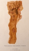
- Pictures
- Online
Dissection of a diseased intestine, showing signs of typhus. Chromolithograph by W. Gummelt, ca. 1897.
Gummelt, W.Date: [1897?]Reference: 577335i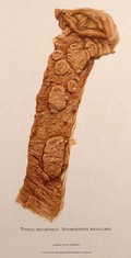
- Pictures
- Online
Dissection of a diseased intestine, showing signs of typhus. Chromolithograph by W. Gummelt, ca. 1897.
Gummelt, W.Date: [1897?]Reference: 577322i- Pictures
Dissection of a man's head and neck showing blood-vessels. Coloured lithograph by William Fairland, 1837, after J. Walsh.
Walsh, J.Date: [1837]Reference: 641914i
- Pictures
- Online
Dissection of the abdomen and thigh of a standing man, showing major blood-vessels. Coloured lithograph by J. Maclise, 1851.
Maclise, Joseph.Date: [1851]Reference: 640789i- Books
Dissection of a recent case of Colles's fracture of the radius : with remarks on the pathology / by John Chiene.
Chiene, John, 1843-1923Date: 1874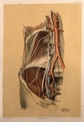
- Pictures
- Online
Dissection of the abdomen of a man, showing the arteries, blood vessels and muscles. Colour lithograph by G.H. Ford, 1866.
Ford, G. H. (George Henry)Date: 1866Reference: 580396i
- Pictures
- Online
Dissection of the abdomen of a man, showing the arteries, veins, and internal organs. Colour lithograph by G.H. Ford, 1865.
Ford, G. H. (George Henry)Date: 1865Reference: 580387i
- Pictures
- Online
Dissection of muscles and blood-vessels of the shoulder and arm of a seated man. Coloured lithograph by J. Maclise, 1851.
Maclise, Joseph.Date: [1851]Reference: 640715i
- Pictures
- Online
Dissection of a portion of the neck, seen from behind: shown resting on a block. Colour lithograph by G.H. Ford, 1865.
Ford, G. H. (George Henry)Date: 1865Reference: 580340i
- Pictures
- Online
Dissection of the abdomen showing the lymphatic vessels. Coloured lithograph by William Fairland, 1837, after W. Bagg after W.J.E. Wilson.
Wilson, Erasmus, Sir, 1809-1884.Date: [1837]Reference: 641996i
- Pictures
- Online
Dissection of the abdomen showing the portal vein. Coloured lithograph by William Fairland, 1837, after J. Walsh after W.J.E. Wilson.
Wilson, Erasmus, Sir, 1809-1884.Date: [1837]Reference: 641995i
- Pictures
- Online
Dissection of a horse's head: seven figures, with details showing the brain and eyeball. Coloured engraving by J. Pass after Harguinier, 1805.
Harguinier, active 1763-1768.Date: 1805Reference: 570688i
- Pictures
- Online
Dissection of a mole: view of the underside of the animal, showing the musculature. Lithograph by E. Wilson after F.W. Brookman, 1880/1900?.
Brookman, F. W.Date: [1880/1900?]Reference: 571431i
- Pictures
- Online
Dissection of a horse, showing the spinal column, head and hind legs, and associated nerves. Coloured engraving by J. Pass after Harguinier, 1805.
Harguinier, active 1763-1768.Date: 1805Reference: 570666i
- Pictures
- Online
Dissection of the area surrounding the anus of a man, with the muscles and blood vessels indicated. Colour lithograph by G.H. Ford, 1865.
Ford, G. H. (George Henry)Date: 1865Reference: 580348i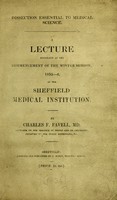
- Books
- Online
Dissection essential to medical science : a lecture delivered at the commencement of the Winter session, 1835-6, at the Sheffield Medical Institution / by Charles F. Favell.
Favell, Charles F.Date: [1836]