Skip to main content
Wellcome Collection homepage
Visit us
What’s on
Stories
Collections
Get involved
About us
Sign in to your library account
Search our stories, images, catalogue and events
Library account
Search our stories, images, catalogue and events
Search
Images search
Search for images
Search
All
Stories
Images
Catalogue
Events
Colours
Licences
Creative Commons CC-BY (18)
Public Domain Mark (15)
Creative Commons CC-BY-NC (14)
In copyright (2)
Dates
From
to
Types/Techniques
Engravings (10)
Manuscript (5)
Etchings (2)
Facsimiles (1)
Lithographs (1)
Photomechanical prints (1)
Prints (1)
Subjects
Drug Industry (3,981)
Pharmaceutical Preparations (3,934)
London (England) (3,872)
AIDS (Disease) (3,026)
Condoms (2,840)
Acquired Immunodeficiency Syndrome (2,557)
HIV Seropositivity (2,513)
Safe Sex (2,216)
Human anatomy (1,842)
HIV Infections - prevention & control (1,756)
Acquired Immunodeficiency Syndrome - prevention & control (1,703)
AIDS (Disease) - Prevention (1,556)
Great Britain (1,549)
Safe sex in AIDS prevention (1,349)
Hospitals (1,284)
HIV Seropositivity - transmission (1,275)
Death (1,273)
Paris (France) (1,130)
ROYAL VETERINARY COLLEGE (1,107)
Charities (1,056)
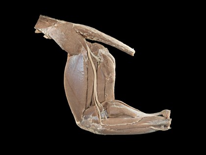
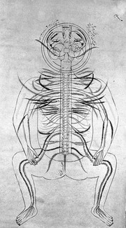
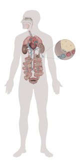
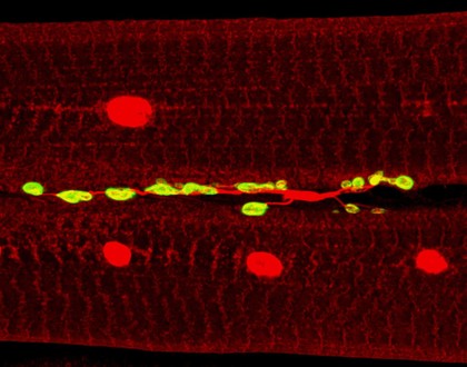
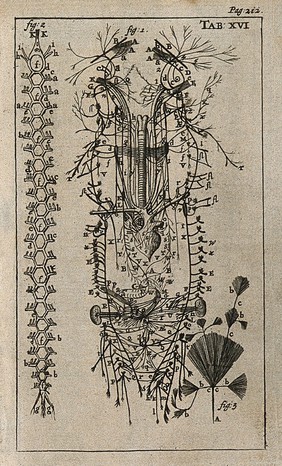
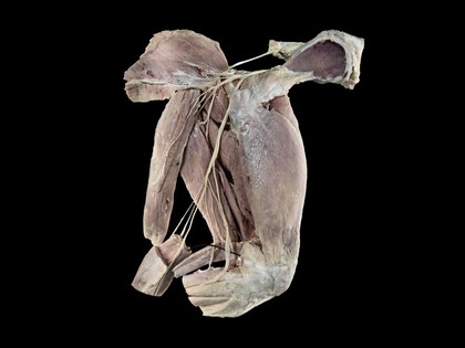
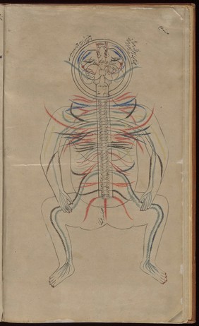
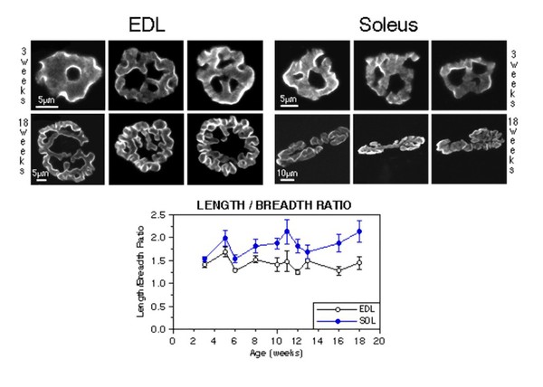
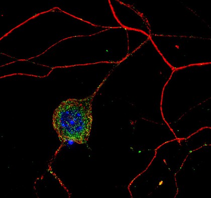
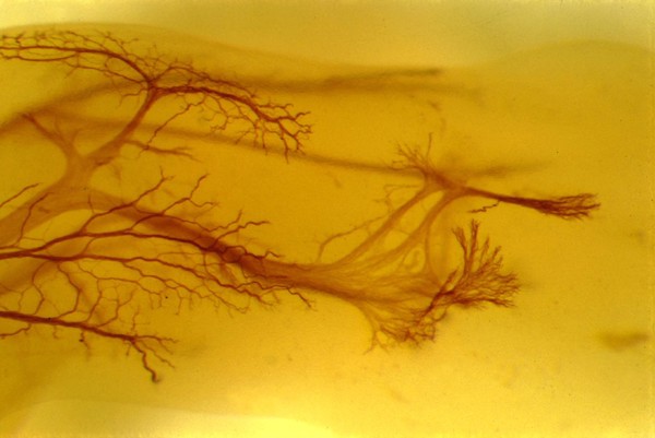
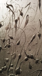
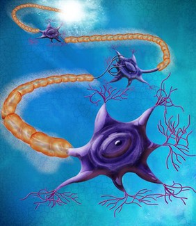
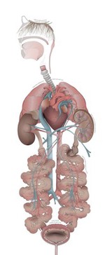
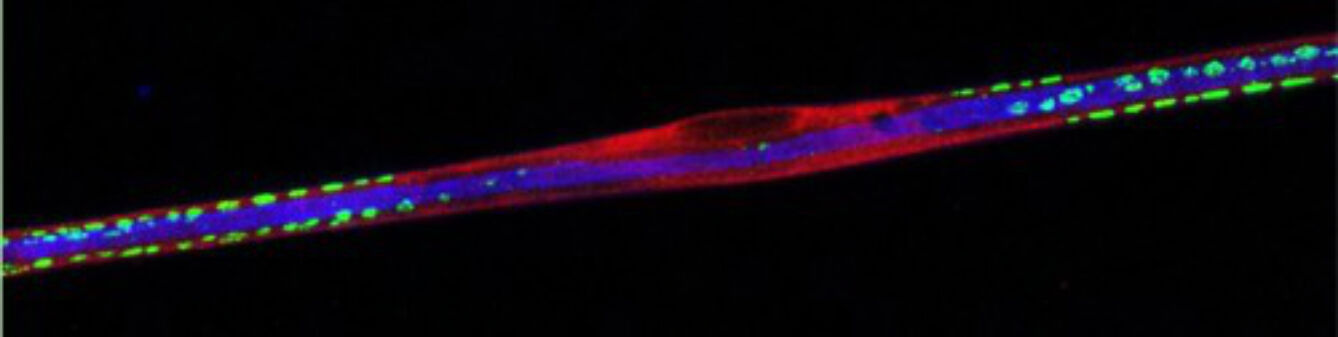
Nerves (49)
Contributors
Dr Guy Bewick, Aberdeen Univ (5)
Dr Jonathan Clarke (4)
Nestor Pestana (4)
Alembert, Jean Le Rond d', 1717-1783 (3)
Diderot, Denis, 1713-1784 (3)
Eustachi, Bartolomeo, -1574 (3)
Michael Frank, Royal Veterinary College (3)
Prof. Peter Brophy (3)
Bénard, 1731-1794 (2)
Dr David Furness (2)
Hermann Aberle, University of Munster (2)
Katja Heuer (2)
Mutlow, H (2)
Vieussens, Raymond, 1641?-1715 (2)
Dr Hermann Aberle, University of Munster (1)
Leonardo, da Vinci, 1452-1519 (1)
Vesalius, Andreas, 1514-1564 (1)
Vesling, Johann, 1598-1649 (1)
Wandelaar, Jan, 1690 or 1692-1759 (1)
Wooding, G (1)
Submit
Active filters:
remove
Nerves
remove
Reset filters
49 results
filtered with: Nerves
Search result sorting
Sort by:
Relevance
Production dates
Sort order:
Ascending
Descending
Submit
Page
1
of 2
Next (page 2)
Close modal window
Page
1
of 2
Next (page 2)