Skip to main content
Wellcome Collection homepage
Visit us
What’s on
Stories
Collections
Get involved
About us
Sign in to your library account
Search our stories, images, catalogue and events
Library account
Search our stories, images, catalogue and events
Search
Images search
Search for images
Search
All
Stories
Images
Catalogue
Events
Colours
Licences
Creative Commons CC-BY (22)
Creative Commons CC-BY-NC (20)
Creative Commons CC0 (1)
Dates
From
to
Types/Techniques
Subjects
Drug Industry (3,981)
Pharmaceutical Preparations (3,935)
London (England) (3,836)
AIDS (Disease) (3,026)
Condoms (2,840)
Acquired Immunodeficiency Syndrome (2,555)
HIV Seropositivity (2,511)
Safe Sex (2,214)
Human anatomy (1,831)
HIV Infections - prevention & control (1,754)
Acquired Immunodeficiency Syndrome - prevention & control (1,703)
AIDS (Disease) - Prevention (1,556)
Great Britain (1,549)
Safe sex in AIDS prevention (1,349)
HIV Seropositivity - transmission (1,275)
Death (1,266)
Hospitals (1,256)
Paris (France) (1,131)
ROYAL VETERINARY COLLEGE (1,107)
Charities (1,056)
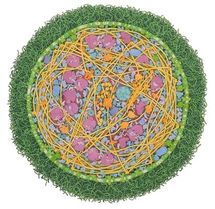
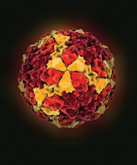
Infectious agent (43)
Contributors
David Goulding, Wellcome Trust Sanger Institute (9)
David S. Goodsell, The Scripps Research Institute (9)
Annie Cavanagh (5)
University of Oxford, Richard Wheeler (4)
Anne Weston, Francis Crick Institute (2)
David S. Goodsell, RCSB Protein Data Bank (2)
Mark Jepson (2)
Michael Frank, Royal Veterinary College (2)
Odra Noel (2)
RCSB Protein Data Bank (2)
Dolores Murcia (1)
Leandro Lemgruber, University of Glasgow (1)
S. Schuller (1)
Stephen Fuller (1)
Submit
Active filters:
remove
Infectious agent
remove
Reset filters
43 results
filtered with: Infectious agent
Search result sorting
Sort by:
Relevance
Production dates
Sort order:
Ascending
Descending
Submit
Page
1
of 2
Next (page 2)
Close modal window
Page
1
of 2
Next (page 2)