Wellcome uses cookies.
Read our policy
Close cookie notification
Skip to main content
Wellcome Collection homepage
Visit us
What’s on
Stories
Collections
Get involved
About us
Sign in to your library account
Search our stories, images, catalogue and events
Library account
Search our stories, images, catalogue and events
Search
Images search
Search for images
Search
All
Stories
Images
Catalogue
Events
Colours
Licences
Creative Commons CC-BY-NC (34)
Creative Commons CC-BY (30)
Creative Commons CC0 (1)
In copyright (1)
Public Domain Mark (1)
Dates
From
to
Types/Techniques
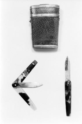
Museum object (4)
Book (1)
Diagrams (1)
Subjects
Drug Industry (3,979)
Pharmaceutical Preparations (3,933)
London (England) (3,818)
AIDS (Disease) (3,026)
Condoms (2,840)
Acquired Immunodeficiency Syndrome (2,555)
HIV Seropositivity (2,511)
Safe Sex (2,214)
Human anatomy (1,830)
HIV Infections - prevention & control (1,754)
Acquired Immunodeficiency Syndrome - prevention & control (1,703)
AIDS (Disease) - Prevention (1,556)
Great Britain (1,549)
Safe sex in AIDS prevention (1,349)
AIDS (1,277)
HIV Seropositivity - transmission (1,275)
Death (1,268)
Hospitals (1,252)
Market (1,235)
AIDS Posters (1,224)
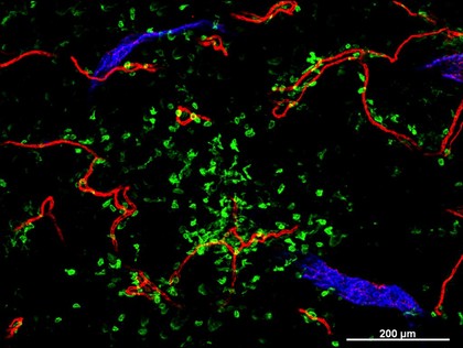
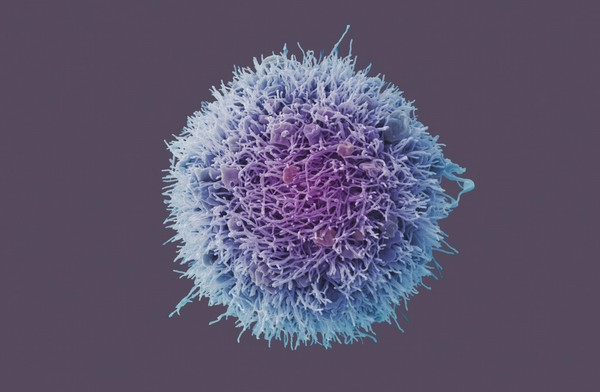
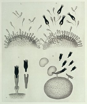
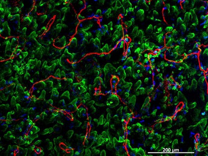
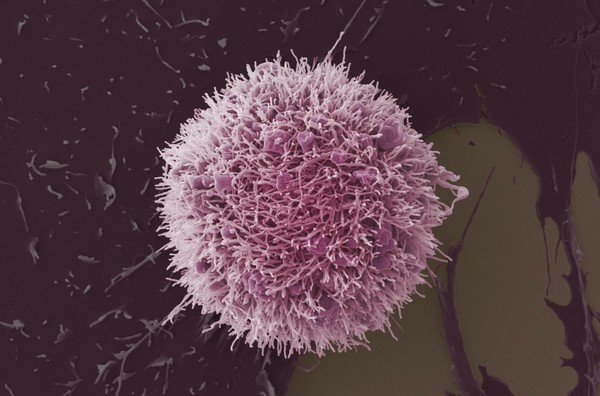
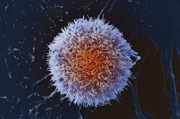
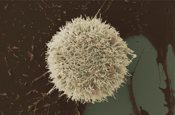
Immunology (67)
Contributors
Anne Weston, Francis Crick Institute (30)
Kevin Mackenzie, University of Aberdeen (12)
Dr. Xiao-nong Wang, Human Dendritic Cell Laboratory, Newcastle University (9)
David Goulding, Wellcome Trust Sanger Institute (4)
Peter Lane and Fiona McConnell (2)
Dolores Murcia (1)
Submit
Active filters:
remove
Immunology
remove
Reset filters
67 results
filtered with: Immunology
Search result sorting
Sort by:
Relevance
Production dates
Sort order:
Ascending
Descending
Submit
Previous (page 1)
Page
2
of 3
Next (page 3)
Close modal window
Previous (page 1)
Page
2
of 3
Next (page 3)