Skip to main content
Wellcome Collection homepage
Visit us
What’s on
Stories
Collections
Get involved
About us
Sign in to your library account
Search our stories, images, catalogue and events
Library account
Search our stories, images, catalogue and events
Search
Images search
Search for images
Search
All
Stories
Images
Catalogue
Events
Colours
Licences
Public Domain Mark (8)
In copyright (1)
Dates
From
to
Types/Techniques
Ephemera (20,093)
Engravings (12,681)
Lithographs (10,506)
Leaflets (10,492)
Portrait prints (9,288)
Photographs (9,169)
Etchings (8,791)
Paintings (6,844)
Posters (5,205)
Photographic prints (5,193)
Watercolors (3,678)
Portrait photographs (3,427)
Prints (2,992)
Wood engravings (2,536)
Oil paintings (2,387)
Stipple engravings (2,269)
Drawings (2,255)
Caricatures (2,116)
Intaglio prints (2,070)
Gouaches (2,017)
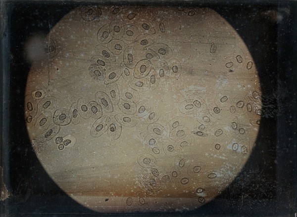
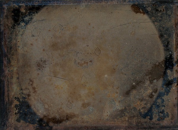
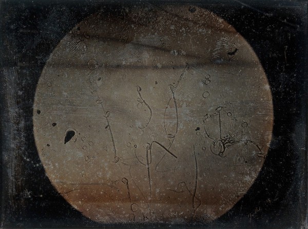
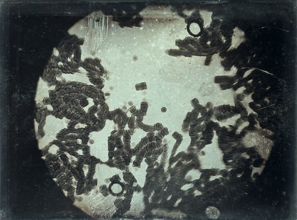
Photomicrographs (13)
Subjects
Kala-azar (1)
Yellow fever (1)
Contributors
Foucault, Léon, 1819-1868 (8)
Submit
Active filters:
remove
Photomicrographs
remove
Reset filters
13 results
filtered with: Photomicrographs
Search result sorting
Sort by:
Relevance
Production dates
Sort order:
Ascending
Descending
Submit
Page
1
of 1
Close modal window
Page
1
of 1