Wellcome uses cookies.
Read our policy
Close cookie notification
Skip to main content
Wellcome Collection homepage
Visit us
What’s on
Stories
Collections
Get involved
About us
Sign in to your library account
Search our stories, images, catalogue and events
Library account
Search our stories, images, catalogue and events
Search
Images search
Search for images
Search
All
Stories
Images
Catalogue
Events
Colours
Licences
Public Domain Mark (30)
In copyright (28)
Creative Commons CC-BY (13)
Creative Commons CC-BY-NC (2)
Dates
From
to
Types/Techniques
Photographic prints (46)
Photographs (42)
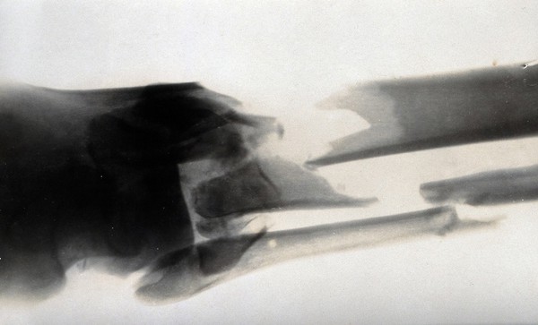
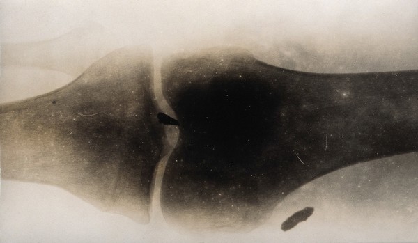
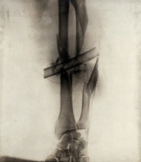
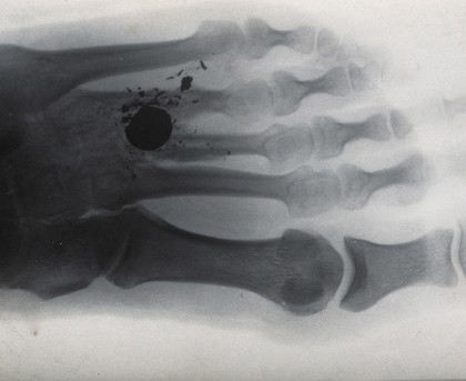
Radiographs (33)
Albumen prints (22)
Gelatin silver prints (15)
Portrait photographs (6)
Electronic journals (1)
Gelatin silver film negatives (1)
Subjects
Cancer (7)
Artwork (6)
Black (6)
Concept (6)
Female (6)
Malignancy (6)
Mammogram (6)
Organ (6)
Preventative medicine (6)
Reproductive system (6)
White (6)
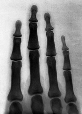
Fingers (3)
Schuster, Arthur, Sir, 1851-1934 (3)
Johannesburg Hospital (2)
Joule, James Prescott, 1818-1889 (2)
Radiation injuries (2)
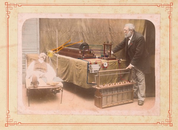
Radiguet, Arthur, 1850-1905 (2)
Radiography (2)
Rings (2)
Schuster, Leonard (2)
Contributors
Röntgen, Wilhelm Conrad, 1845-1923 (11)
Wimshurst, James, 1832-1903 (7)
Chiara Salvi (6)
Melhuish, Arthur James, 1829-1895 (3)
Schuster, Arthur, Sir, 1851-1934 (3)
Radiguet, Arthur, 1850-1905 (2)
Radiographie Radiguet (2)
Ropner, L., active 1897 (2)
Roscoe, Lucy, -1910 (2)
Sutcliffe, Frank Meadow, 1853-1941 (2)
Finzi, Neville Samuel, 1881-1968 (1)
Hrdlička, Aleš, 1869-1943 (1)
Manson-Bahr, Philip H. (Philip Henry), Sir, 1881-1966 (1)
Newton & Wright (photographers) (1)
Orton, G. Harrison (George Harrison), 1873- (1)
Shuttleworth, G. E. (George Edward), 1842-1928 (1)
Wood, F (1)
Submit
96 results
Search result sorting
Sort by:
Relevance
Production dates
Sort order:
Ascending
Descending
Submit
Previous (page 1)
Page
2
of 4
Next (page 3)
Close modal window
Previous (page 1)
Page
2
of 4
Next (page 3)