Wellcome uses cookies.
Read our policy
Close cookie notification
Skip to main content
Wellcome Collection homepage
Visit us
What’s on
Stories
Collections
Get involved
About us
Sign in to your library account
Search our stories, images, catalogue and events
Library account
Search our stories, images, catalogue and events
Search
Images search
Search for images
Search
All
Stories
Images
Catalogue
Events
Colours
Licences
Public Domain Mark (64)
Creative Commons CC-BY-NC (4)
In copyright (2)
Dates
From
to
Types/Techniques
Engravings (41)
Intaglio prints (16)
Etchings (8)
Title pages (3)
Lithographs (2)
Posters (2)
Photographs (1)
Subjects
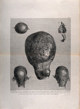
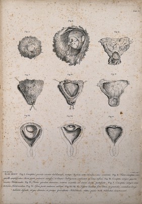
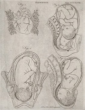
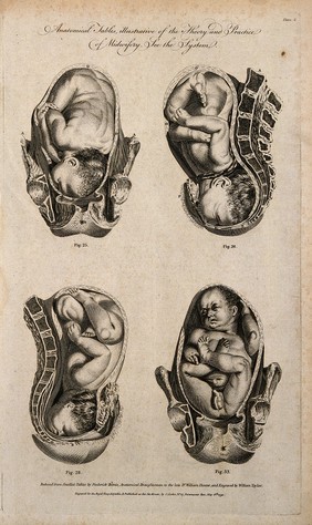
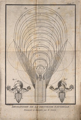
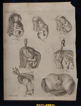
Uterus, Pregnant (68)
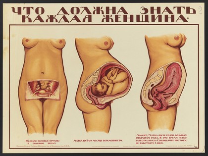
Pregnancy (58)
Human anatomy (42)
Uterus - anatomy & histology (35)
Gynecologic pathology (21)
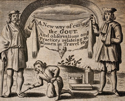
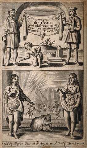
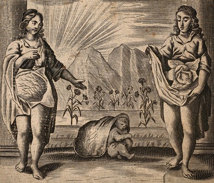
Childbirth (18)
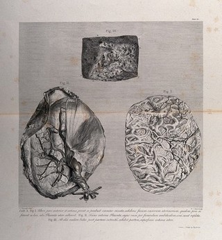
Placenta (18)
Obstetrics (9)
Labor (Obstetrics) - Complications (8)
Breech delivery (7)
Delivery (Obstetrics) (7)
Obstetrical forceps (7)
Human dissection (6)
Twins (6)
Fetus - Growth (5)
Pelvis (5)
Anatomy (3)
Development (3)
Fetus - Anatomy (3)
Generative organs, Female (3)
Contributors
Hunter, William, 1718-1783 (39)
Rymsdyk, Jan van, active 1750-1788 (33)
Bell, Andrew, 1726-1809 (10)
Menil (6)
Smellie, William, 1697-1763 (6)
Birnie, Frederick (5)
Hall, William Henry, -1807 (5)
Aliamet, François Germain, 1734-1788 (4)
Canot, Pierre Charles, 1710-1777 (4)
Barlow, Inigo (3)
Michael Frank, Royal Veterinary College (3)
Taylor, J (3)
Gri︠u︡n, O., active 1919 (2)
Levret, A. (André), 1703-1780 (2)
Müller, J. S (2)
Ravenet, Simon François, 1706-1774 (2)
Scotin, active 1765 (2)
Scotin, Gérard, the elder, 1643-1715 (2)
Soviet Union. Otdel okhrany materinstva i mladenchestva (2)
Taylor, William, active 1791 (2)
Submit
73 results
Search result sorting
Sort by:
Relevance
Production dates
Sort order:
Ascending
Descending
Submit
Previous (page 1)
Page
2
of 3
Next (page 3)
Close modal window
Previous (page 1)
Page
2
of 3
Next (page 3)