Skip to main content
Wellcome Collection homepage
Visit us
What’s on
Stories
Collections
Get involved
About us
Sign in to your library account
Search our stories, images, catalogue and events
Library account
Search our stories, images, catalogue and events
Search
Images search
Search for images
Search
All
Stories
Images
Catalogue
Events
Colours
Licences
Public Domain Mark (33)
In copyright (23)
Creative Commons CC-BY (2)
Dates
From
to
Types/Techniques
Photographs (51)
Albumen prints (39)
Photomechanical prints (23)
Posters (23)
Protest posters (23)
Gelatin silver prints (11)
Portrait photographs (5)
Caricatures (3)
Paintings (3)
Photographic prints (3)
Portrait paintings (3)
Oil paintings (2)
Woodcuts (2)
Acrylic paintings (1)
Engravings (1)
Group portraits (1)
Lithographs (1)
Watercolors (1)
Subjects
Abortion - Moral and ethical aspects (23)
Fetus (23)
Great Britain (23)
Pro-choice movement (23)
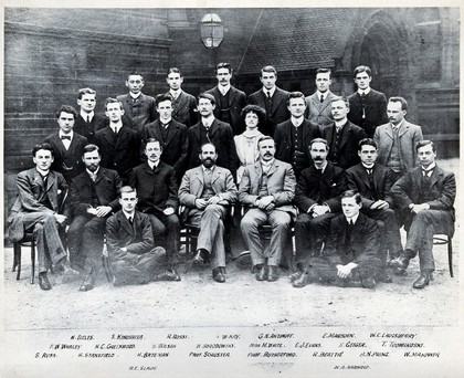
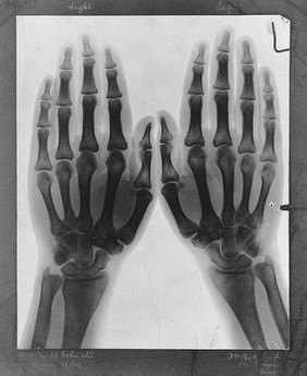
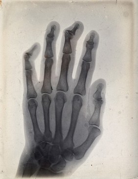
Schuster, Arthur, Sir, 1851-1934 (4)
Joule, James Prescott, 1818-1889 (2)
Schuster, Leonard (2)
Schuster, Norah Henriette, 1892-1991 (2)
Addison, Joseph, 1672-1719 (1)
Blythswood, Archibald Campbell Campbell, Baron, 1835-1908 (1)
Bridget, of Sweden, Saint, approximately 1303-1373 (1)
Christopher, Saint (1)
COWS (1)
Death (1)
Dexter, T. Michael, 1945- (1)
George III, King of Great Britain, 1738-1820 (1)
George IV, King of Great Britain, 1762-1830 (1)
Gladstone, W. E. (William Ewart), 1809-1898 (1)
Goodwin, Anthony, -1819 (1)
Great Britain. Parliament (1)
Contributors
Schuster, Arthur, Sir, 1851-1934 (32)
Melhuish, Arthur James, 1829-1895 (3)
Ottley, William Young, 1771-1836 (2)
Roscoe, Lucy, -1910 (2)
Sutcliffe, Frank Meadow, 1853-1941 (2)
Bonnar, W. (William), 1800-1853 (1)
Dexter, Alex (Alexander Michael), 1972- (1)
Rawlinson, James, 1769-1848 (1)
Rowlandson, Thomas, 1756-1827 (1)
Submit
83 results
Search result sorting
Sort by:
Relevance
Production dates
Sort order:
Ascending
Descending
Submit
Page
1
of 3
Next (page 2)
Close modal window
Page
1
of 3
Next (page 2)