Wellcome uses cookies.
Read our policy
Close cookie notification
Skip to main content
Wellcome Collection homepage
Visit us
What’s on
Stories
Collections
Get involved
About us
Sign in to your library account
Search our stories, images, catalogue and events
Library account
Search our stories, images, catalogue and events
Search
Images search
Search for images
Search
All
Stories
Images
Catalogue
Events
Colours
Licences
Public Domain Mark (275)
Creative Commons CC-BY (119)
In copyright (21)
Creative Commons CC-BY-NC (10)
Creative Commons CC0 (3)
Dates
From
to
Types/Techniques
Book illustrations (32)
Engravings (31)
Wood engravings (30)
Etchings (20)
Photographic prints (18)
Lithographs (15)
Paintings (14)
Photographs (13)
Watercolors (11)
Ephemera (9)
Landscape drawings (8)
Photograph albums (8)
Portrait prints (8)
Drawings (6)
Posters (6)
Prints (6)
Incunabula (5)
Gouaches (4)
Line photoengravings (4)
Monogrammed bindings (Binding) (4)
Subjects
Epilepsy (46)
Hypnosis (46)
Hysteria (46)
Photography (46)
Surgical Instruments (43)
Anatomy (37)
Food groups (29)
Healthy eating (29)
Human anatomy (29)
Nutrients (29)
Nutrition (29)
Cut out (28)
Pattern (27)
General Surgery (24)
Cancer (20)
White (19)
Spine (18)
Structure (18)
3D (17)
3D printing (17)
Contributors
Birmingham Medical Institute (46)
Bourneville, 1840-1909 (46)
Charcot, J. M. (Jean Martin), 1825-1893 (46)
Regnard, Paul, 1850-1927 (46)
Salpêtrière (Hospital) (46)
Paré, Ambroise, 1510?-1590 (41)
Alexandr Khrapichev, University of Oxford (29)
Rasse Desneux, Nicolas, -1581 (27)
Dave Farnham (16)
Medical Society of London (15)
Bell, Charles, Sir, 1774-1842 (14)
Royal Humane Society (London, England) (12)
Foot, Jesse, 1744-1826 (10)
Bodenstein, Adam von, 1528-1577 (9)
British Library (9)
Howard, John, 1726-1790 (9)
Paracelsus, 1493-1541 (9)
Sayre, Lewis A. (Lewis Albert), 1820-1900 (9)
Cesnola, Alessandro Palma di, 1839-1914 (8)
Mercuriale, Girolamo, 1530-1606 (7)
Submit
624 results
Search result sorting
Sort by:
Relevance
Production dates
Sort order:
Ascending
Descending
Submit
Previous (page 1)
Page
2
of 21
Next (page 3)
Close modal window
Previous (page 1)
Page
2
of 21
Next (page 3)
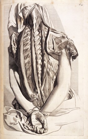


![Observations on injuries of the spine and of the thigh bone: in two lectures. Delivered in the school of Great Windmill street / [Sir Charles Bell].](https://iiif.wellcomecollection.org/image/L0009923/full/282%2C/0/default.jpg)
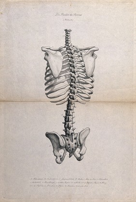
![Observations on injuries of the spine and of the thigh bone: in two lectures. Delivered in the school of Great Windmill street / [Sir Charles Bell].](https://iiif.wellcomecollection.org/image/L0063650/full/282%2C/0/default.jpg)
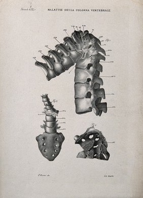
![Observations on injuries of the spine and of the thigh bone: in two lectures. Delivered in the school of Great Windmill street / [Sir Charles Bell].](https://iiif.wellcomecollection.org/image/L0023984/full/282%2C/0/default.jpg)
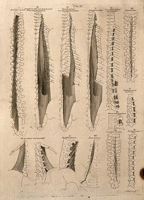
![Observations on injuries of the spine and of the thigh bone: in two lectures. Delivered in the school of Great Windmill street / [Sir Charles Bell].](https://iiif.wellcomecollection.org/image/L0009924/full/320%2C/0/default.jpg)