Wellcome uses cookies.
Read our policy
Close cookie notification
Skip to main content
Wellcome Collection homepage
Visit us
What’s on
Stories
Collections
Get involved
About us
Sign in to your library account
Search our stories, images, catalogue and events
Library account
Search our stories, images, catalogue and events
Search
Images search
Search for images
Search
All
Stories
Images
Catalogue
Events
Colours
Licences
Public Domain Mark (15)
Creative Commons CC-BY (6)
In copyright (3)
Creative Commons CC-BY-NC (1)
Dates
From
to
Types/Techniques
Book illustrations (9)
Lithographs (3)
Drawings (2)
Etchings (2)
Ink drawings (2)
Photographic prints (2)
Advertisements (1)
Engravings (1)
Facsimiles (1)
Motion picture posters (1)
Photomechanical prints (1)
Posters (1)
Prints (1)
Subjects
Channels (6)
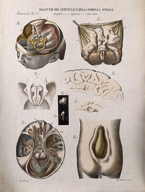
Human anatomy (6)
Acu-moxa (5)
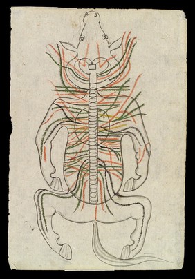
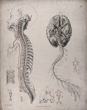
Spine (4)
Measurement (3)
Nerves (3)
Proportionate body measurement (3)
Blood - Circulation (2)
Brain (2)
Ming period (1368-1644) (2)
Pasteur Institute of India, Kasauli (2)
Rabies (2)
Spinal column (2)
Acupuncture chart (1)
Backbone (1)
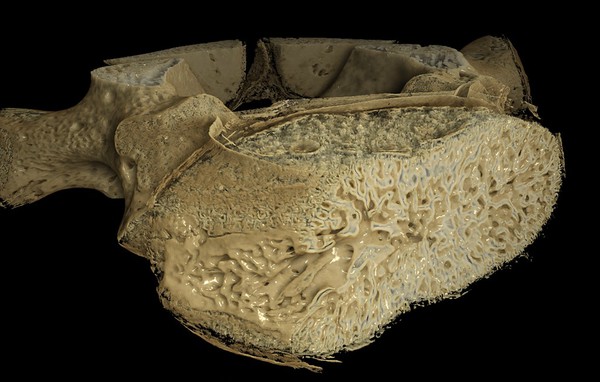
Bone and Bones (1)
Bones (1)
Bronze figure (1)
Chinese Medicine (1)
Coal mines and mining (1)
Contributors
Acquaah, Frank (1)
Basire, Isaac, 1704-1768 (1)
Batelli (1)
Dr Steve Wilson (1)
Eustachi, Bartolomeo, -1574 (1)
Evelyn, John, 1620-1706 (1)
Goya, Francisco, 1746-1828 (1)
Gucht, Michael van der, 1660-1725 (1)
Harguinier, active 1763-1768 (1)
James, R. (Robert), 1703?-1776 (1)
Luo Shaoji (1)
Maclise, Joseph (1)
Muzzi, Ottavio (1)
Pass, J (1)
Progress Film-Verleih (Berlin, Germany) (1)
Vesling, Johann, 1598-1649 (1)
Vigevano, Guido da, approximately 1280-approximately 1350 (1)
Wellcome Images (1)
Wongel (Graphic designer), active approximately 1976- (1)
Żeromski, Stefan, 1864-1925 (1)
Submit
27 results
Search result sorting
Sort by:
Relevance
Production dates
Sort order:
Ascending
Descending
Submit
Page
1
of 1
Close modal window
Page
1
of 1