Wellcome uses cookies.
Read our policy
Close cookie notification
Skip to main content
Wellcome Collection homepage
Visit us
What’s on
Stories
Collections
Get involved
About us
Sign in to your library account
Search our stories, images, catalogue and events
Library account
Search our stories, images, catalogue and events
Search
Images search
Search for images
Search
All
Stories
Images
Catalogue
Events
Colours
Licences
Public Domain Mark (66)
Creative Commons CC-BY-NC (2)
In copyright (1)
Dates
From
to
Types/Techniques
Photographs (62)
Albumen prints (43)
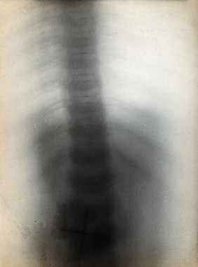
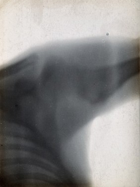
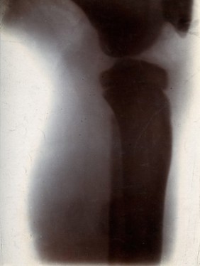
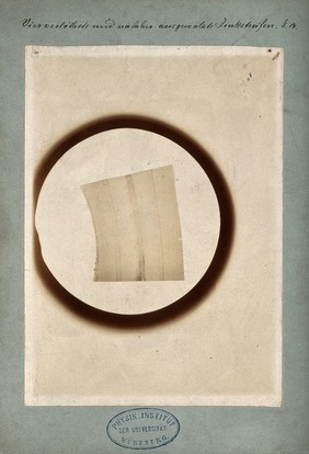
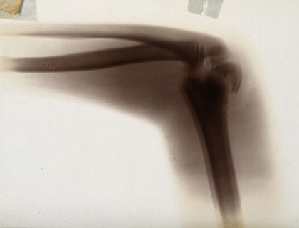
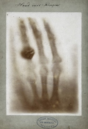
Radiographs (29)
Photographic prints (23)
Gelatin silver prints (8)
Lithographs (7)
Floor plans (1)
Group portraits (1)
Photolithographs (1)
Prints (1)
Silver gelatin photoprints (1)
Subjects
Hood, Basil, 1876-1978 (8)
Africa (7)
Explorers (7)
Livingstone, David, 1813-1873 (7)
Missionaries (7)
St. Charles Hospital (London, England) (5)
Bézu St. Eloi (Eure, France) (2)
Brongniart, Alexandre, 1770-1847 (2)
Nurses (2)
St. Marylebone Infirmary (London, England) (2)
Tennis (2)
Ailly, A., active 1790-1818 (1)
Alison, William Pulteney, 1790-1859 (1)
Ampère, André-Marie, 1775-1836 (1)
Apothecary jars - Netherlands - 19th century (1)
Ballingall, George, 1780-1855 (1)
Bucknall (England) (1)
Christianity (1)
Christison, Robert, Sir, bart., 1797-1882 (1)
Collège de France (Paris, France) (1)
Contributors
Schuster, Arthur, Sir, 1851-1934 (32)
Wimshurst, James, 1832-1903 (18)
Röntgen, Wilhelm Conrad, 1845-1923 (11)
Brongniart, Alexandre, 1770-1847 (1)
Jelgerhuis, J. (Johannes), 1770-1836 (1)
Jones, Elijah, active 1884-1902 (1)
Sprague & Co. (London, England) (1)
V.A. Bruckmann (1904 : Munich) (1)
Submit
83 results
Search result sorting
Sort by:
Relevance
Production dates
Sort order:
Ascending
Descending
Submit
Previous (page 1)
Page
2
of 3
Next (page 3)
Close modal window
Previous (page 1)
Page
2
of 3
Next (page 3)