Wellcome uses cookies.
Read our policy
Close cookie notification
Skip to main content
Wellcome Collection homepage
Visit us
What’s on
Stories
Collections
Get involved
About us
Sign in to your library account
Search our stories, images, catalogue and events
Library account
Search our stories, images, catalogue and events
Search
Images search
Search for images
Search
All
Stories
Images
Catalogue
Events
Colours
Licences
Creative Commons CC-BY (31)
Creative Commons CC-BY-NC (14)
Creative Commons CC0 (1)
Dates
From
to
Types/Techniques
Subjects
Leucocyte (30)
White blood cell (20)
Blue (15)
Immunology (14)
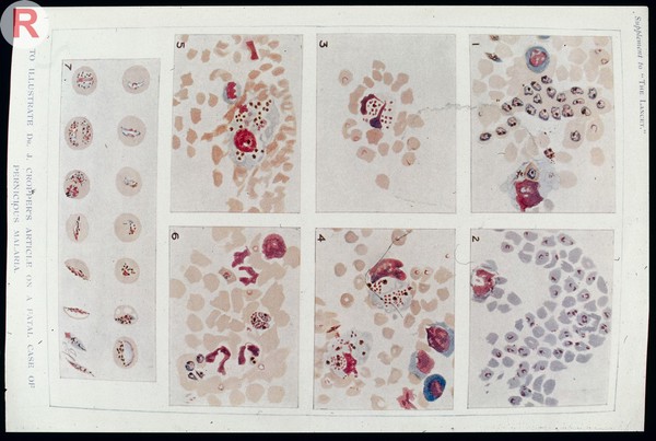
Red (14)
White blood cells (13)
Green (11)
Immune response (10)
Red blood cells (10)
Barriers (9)
Clusters (9)
Connections (9)
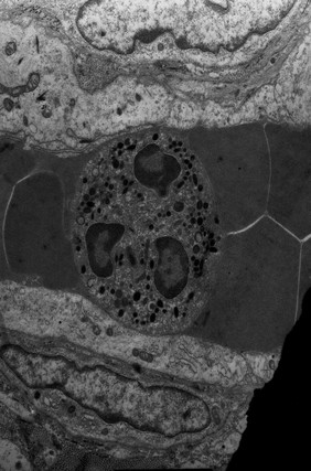
Electron micrographs (9)
ELECTRON MICROSCOPE (9)
Network (9)
Protection (9)
T cell (9)
Wholemount (9)
Clot (8)
Healing (8)
Contributors
David Gregory & Debbie Marshall (9)
Dr. Xiao-nong Wang, Human Dendritic Cell Laboratory, Newcastle University (9)
Rob Young (9)
Kevin Mackenzie, University of Aberdeen (6)
University of Edinburgh (5)
S. Gschmeissner, K. Hodivala-Dilke & M. Stone (2)
A. Walker, L. Sharp & J. Pryde (1)
Mateus Crespo, The Institute of Cancer Research (1)
Neil Dufton (1)
Submit
46 results
Search result sorting
Sort by:
Relevance
Production dates
Sort order:
Ascending
Descending
Submit
Previous (page 1)
Page
2
of 2
Close modal window
Previous (page 1)
Page
2
of 2