Wellcome uses cookies.
Read our policy
Close cookie notification
Skip to main content
Wellcome Collection homepage
Visit us
What’s on
Stories
Collections
Get involved
About us
Sign in to your library account
Search our stories, images, catalogue and events
Library account
Search our stories, images, catalogue and events
Search
Images search
Search for images
Search
All
Stories
Images
Catalogue
Events
Colours
Licences
Public Domain Mark (10)
Dates
From
to
Types/Techniques
Etchings (7)
Engravings (3)
Subjects
Human anatomy (10)
Blood - Circulation (3)
Brain (3)
Nerves (3)
Muscles (2)
Anatomy, Artistic (1)
Bones (1)
Diaphragm (1)
Drawing (1)
Generative organs, Female (1)
Genitourinary organs (1)
Human skeleton (1)
Larynx (1)
Pharynx (1)
Skull (1)
Spine (1)
Testis (1)
Contributors
Eustachi, Bartolomeo, -1574 (10)
James, R. (Robert), 1703?-1776 (7)
Lancisi, Giovanni Maria, 1654-1720 (7)
Basire, Isaac, 1704-1768 (4)
Alembert, Jean Le Rond d', 1717-1783 (3)
Diderot, Denis, 1713-1784 (3)
Bénard, 1731-1794 (2)
Cowper, William, 1666-1709 (2)
Gucht, Michael van der, 1660-1725 (2)
Vieussens, Raymond, 1641?-1715 (2)
Accademia di Francia (Rome, Italy) (1)
Bickham, George, 1684?-1758? (1)
Cheselden, William, 1688-1752 (1)
Duverney, M. (Jacques-François-Marie), 1661-1748 (1)
Evelyn, John, 1620-1706 (1)
Graaf, Reinier de, 1641-1673 (1)
Haller, Albrecht von, 1708-1777 (1)
Senex, John, -1740 (1)
Van der Gucht, Gerard, 1696-1776 (1)
Vesling, Johann, 1598-1649 (1)
Submit
11 results
Search result sorting
Sort by:
Relevance
Production dates
Sort order:
Ascending
Descending
Submit
Page
1
of 1
Close modal window
Page
1
of 1
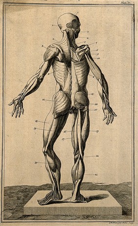
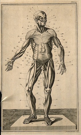
![Anatomy improv'd and illustrated with regard to the uses thereof in designing: not only laid down from an examen of the bones and muscles of the human body, but also demonstrated and exemplified from the most celebrated antique statues in Rome. Exhibited in a great number of copper plates, with all the figures in various views / Intended originally for y use of the Royal French Academy of Painting and Sculpture. And carried on under the care and inspection of Charles Errard director of the same in Rome. The dissections made by Doc[to]r Ber[nardin]o Genga ... The explanations and indexes added by ... John Maria Lancissi ... First published at Rome by Dom di Rossi and now reengraven ... And republish'd by John Senex. A work of great use to painters, sculptors, statuaries and all others studious in the noble art of designing.](https://iiif.wellcomecollection.org/image/M0018007/full/180%2C/0/default.jpg)