Wellcome uses cookies.
Read our policy
Close cookie notification
Skip to main content
Wellcome Collection homepage
Visit us
What’s on
Stories
Collections
Get involved
About us
Sign in to your library account
Search our stories, images, catalogue and events
Library account
Search our stories, images, catalogue and events
Search
Images search
Search for images
Search
All
Stories
Images
Catalogue
Events
Colours
Licences
Creative Commons CC-BY (71)
Public Domain Mark (2)
Dates
From
to
Types/Techniques
Subjects
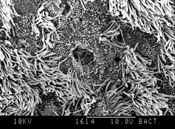
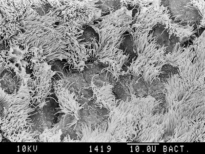
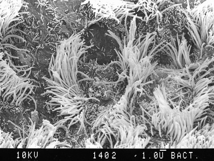
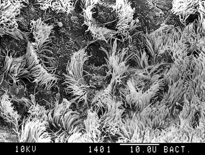
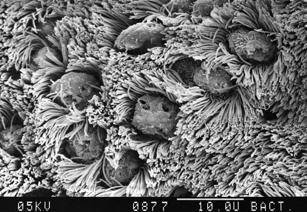
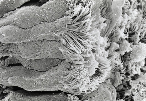
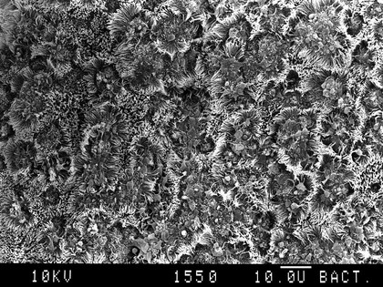
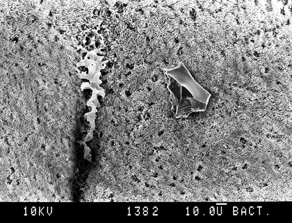
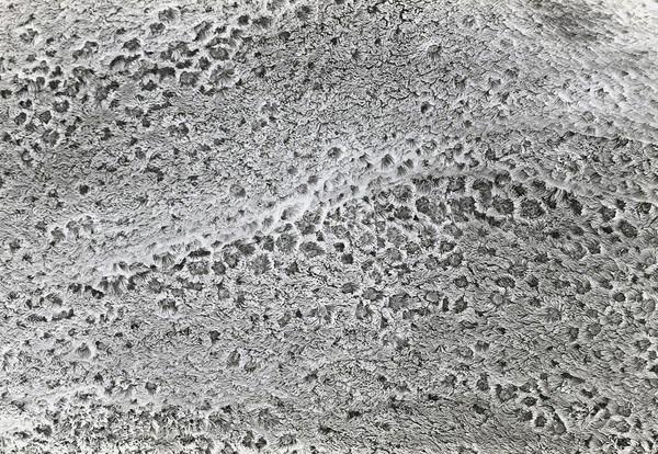
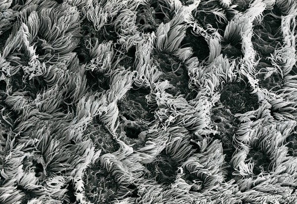
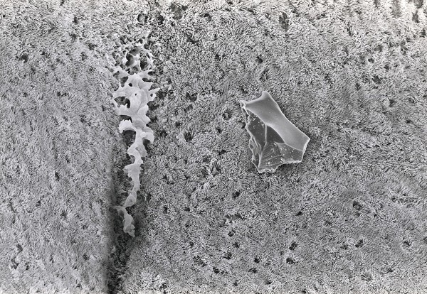
BRONCHUS (38)
SEM (36)
SCANNING ELECTRON MICROSCOPE (30)
Bronchus (25)
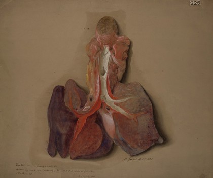
Lungs (22)
Breathing (21)
Electron micrographs (17)
3D (15)
STEREOSCOPIC IMAGE (15)
Human (14)
HUMAN (12)
GOBLET CELL (8)
Lung function (8)
EMPHYSAEMA (4)
ALVEOLI (3)
CILIATED EPITHELIUM (3)
COLUMNAR EPITHELIUM (3)
Goblet cell (3)
Normal (3)
NORMAL (3)
Contributors
David Gregory & Debbie Marshall (61)
David Gregory and Debbie Marshall (6)
Godart, Thomas (2)
John George Adami (2)
Submit
73 results
Search result sorting
Sort by:
Relevance
Production dates
Sort order:
Ascending
Descending
Submit
Previous (page 1)
Page
2
of 3
Next (page 3)
Close modal window
Previous (page 1)
Page
2
of 3
Next (page 3)