Wellcome uses cookies.
Read our policy
Close cookie notification
Skip to main content
Wellcome Collection homepage
Visit us
What’s on
Stories
Collections
Get involved
About us
Sign in to your library account
Search our stories, images, catalogue and events
Library account
Search our stories, images, catalogue and events
Search
Images search
Search for images
Search
All
Stories
Images
Catalogue
Events
Colours
Licences
Public Domain Mark (34)
Creative Commons CC-BY (8)
Creative Commons CC-BY-NC (2)
Dates
From
to
Types/Techniques
Photographs (62)
Albumen prints (50)
Gelatin silver prints (11)
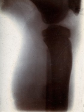
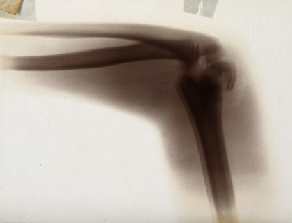
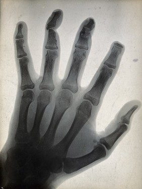
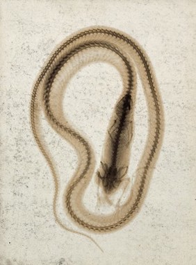
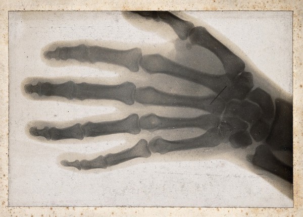
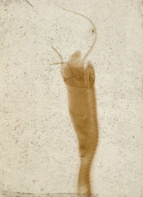
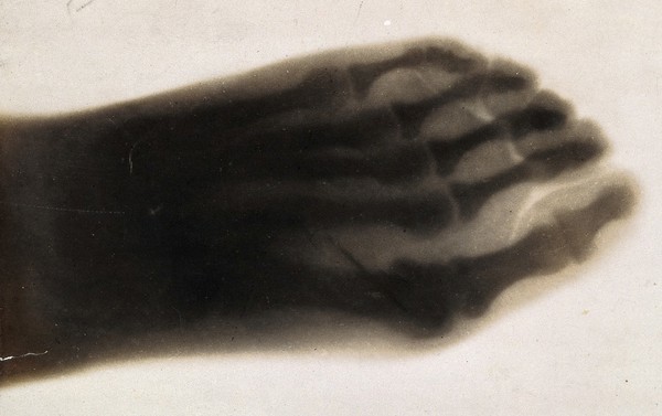
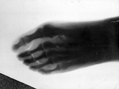
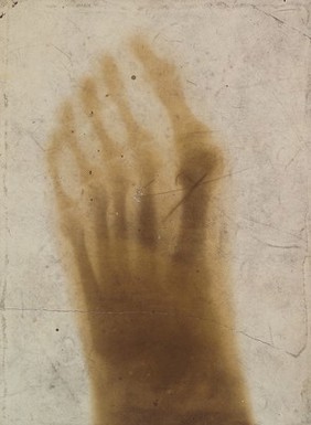
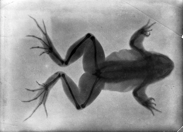
Radiographs (11)
Portrait photographs (5)
Photographic prints (3)
Drawings (1)
Group portraits (1)
Ink drawings (1)
Subjects
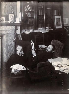
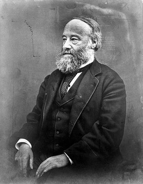
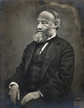
Schuster, Arthur, Sir, 1851-1934 (5)
Joule, James Prescott, 1818-1889 (2)
Schuster, Leonard (2)
Schuster, Norah Henriette, 1892-1991 (2)
Balfour, Arthur James, 1848-1930 (1)
Blanc, G. A. (Gian Alberto) (1)
Blythswood, Archibald Campbell Campbell, Baron, 1835-1908 (1)
British Association for the Advancement of Science (1)
Cambridge (England) (1)
Elster, Julius, 1854-1920 (1)
Geitel, Hans Friedrich Karl, 1855-1923 (1)
Kelvin, William Thomson, Baron, 1824-1907 (1)
Lamb, Horace, Sir, 1849-1934 (1)
Lodge, Oliver, Sir, 1851-1940 (1)
Physics - Education (1)
Rayleigh, John William Strutt, Baron, 1842-1919 (1)
Roentgen rays (1)
Roentgen Rays (1)
Rutherford, Ernest, 1871-1937 (1)
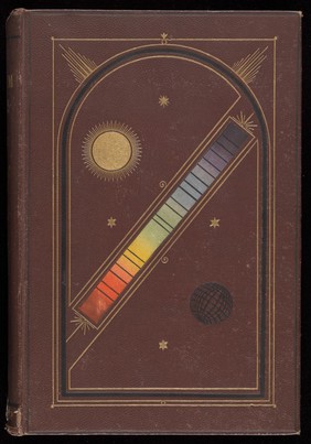
Spectrum Analysis (1)
Contributors
Schuster, Arthur, Sir, 1851-1934 (33)
Röntgen, Wilhelm Conrad, 1845-1923 (11)
Melhuish, Arthur James, 1829-1895 (3)
Arthur Schuster (2)
Grimmett of Banbury (2)
Roscoe, Lucy, -1910 (2)
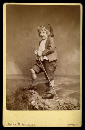
Sutcliffe, Frank Meadow, 1853-1941 (2)
Boyd, A. S. (Alexander Stuart), 1854-1930 (1)
Elliott & Fry (1)
Lafayette Ltd (1)
Roscoe, Henry E. (Henry Enfield), 1833-1915 (1)
Submit
72 results
Search result sorting
Sort by:
Relevance
Production dates
Sort order:
Ascending
Descending
Submit
Previous (page 1)
Page
2
of 3
Next (page 3)
Close modal window
Previous (page 1)
Page
2
of 3
Next (page 3)