Wellcome uses cookies.
Read our policy
Close cookie notification
Skip to main content
Wellcome Collection homepage
Visit us
What’s on
Stories
Collections
Get involved
About us
Sign in to your library account
Search our stories, images, catalogue and events
Library account
Search our stories, images, catalogue and events
Search
Images search
Search for images
Search
All
Stories
Images
Catalogue
Events
Colours
Licences
Public Domain Mark (105)
Creative Commons CC-BY (2)
Creative Commons CC-BY-NC (2)
In copyright (1)
Dates
From
to
Types/Techniques
Photographs (77)
Albumen prints (45)
Lithographs (26)
Caricatures (21)
Wet collodion negatives (16)
Gelatin silver prints (15)
Paintings (14)
Watercolors (14)
Portrait paintings (11)
Portrait prints (11)
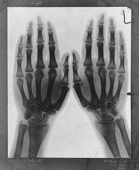
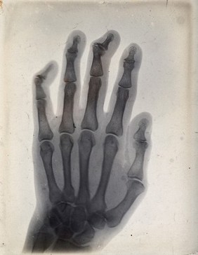
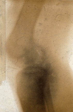
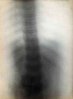
Radiographs (11)
Cityscape photographs (8)
Mezzotints (8)
Group portraits (6)
Oil paintings (5)
Portrait photographs (5)
Etchings (4)
Waterscape photographs (4)
Photomechanical prints (3)
Prints (3)
Subjects
Hong Kong (China) (29)
Skin - Diseases (12)
Wette, Ludwig de, 1812-1887 (5)
Ancient (3)
Archaeology (3)
Eyeglasses (3)
Hospitals - England (3)
Jews - history (3)
Lachish (Israel) (3)
Paleography, Hebrew (3)
Palestine (3)
Sick (3)
Wellington, Arthur Wellesley, Duke of, 1769-1852 (3)
Alfred, Duke of Saxe-Coburg and Gotha, 1844-1900 (2)
Architecture - England (2)
Barcelona (Spain) (2)
Blind (2)
Blind - Books and reading - Great Britain (2)
Blind - Services for (2)
Cayley, Arthur, 1821-1895 (2)
Contributors
Schuster, Arthur, Sir, 1851-1934 (32)
Thomson, J. (John), 1837-1921 (29)
Doyle, John, 1797-1868 (20)
Hutchinson, Jonathan, Sir, 1828-1913 (13)
Green, Mabel (12)
Röntgen, Wilhelm Conrad, 1845-1923 (11)
Wette-Jersing, Amalia de, 1816-1898 (5)
Say, William, 1768-1834 (3)
Starkey, J. L. (James Leslie), 1895-1938 (3)
Tufnell, Olga (3)
Tur-Sinai, Naphtali H. (Naphtali Herz), 1886-1973 (3)
Ward, William, 1766-1826 (3)
Casas, Ramón, 1866-1932 (2)
Draycott, John A. (Photographer), active approximately 1896 (2)
Lorimer, John Henry, 1856-1936 (2)
Meyer, H. (Henri), 1841-1899 (2)
Sanatorio para sifilíticos (2)
Smith, John Raphael, 1752-1812 (2)
Thomàs i Bigas, Josep, 1852-1910 (2)
Thorne, Diana, 1895-1963 (2)
Submit
162 results
Search result sorting
Sort by:
Relevance
Production dates
Sort order:
Ascending
Descending
Submit
Previous (page 2)
Page
3
of 6
Next (page 4)
Close modal window
Previous (page 2)
Page
3
of 6
Next (page 4)