Wellcome uses cookies.
Read our policy
Close cookie notification
Skip to main content
Wellcome Collection homepage
Visit us
What’s on
Stories
Collections
Get involved
About us
Sign in to your library account
Search our stories, images, catalogue and events
Library account
Search our stories, images, catalogue and events
Search
Images search
Search for images
Search
All
Stories
Images
Catalogue
Events
Colours
Licences
Public Domain Mark (2,375)
Creative Commons CC-BY-NC (424)
Creative Commons CC-BY (401)
In copyright (381)
Creative Commons CC0 (6)
Creative Commons CC-BY-NC-ND (2)
Dates
From
to
Types/Techniques
Paintings (992)
Oil paintings (842)
Engravings (593)
Photographs (574)
Lithographs (495)
Portrait paintings (405)
Wet collodion negatives (297)
Etchings (237)
Watercolors (234)
Photographic prints (230)
Posters (192)
Photograph albums (140)
Portrait prints (135)
Caricatures (131)
Ephemera (110)
Prints (100)
Drawings (97)
Genre photographs (78)
Photomechanical prints (74)
Leaflets (64)
Subjects
Human anatomy (304)
Cyprus (140)
Blood - Circulation (112)
Fungi - England (106)
London (England) (102)
Botany - England - History - 19th century (99)
AIDS (Disease) (98)
Mycology - England - History - 19th century (96)
Anatomy (92)
Pechili Province (China) (91)
Mushrooms (87)
Cook, James, 1728-1779 Travel (80)
China (74)
Guangdong Sheng (China) (70)
Women (67)
Death (64)
Fujian Sheng (China) (64)
Identity (Psychology) in art (61)
Art and mental illness (60)
Schizophrenia and the arts (60)
Contributors
Thomson, J. (John), 1837-1921 (300)
Cesnola, Alessandro Palma di, 1839-1914 (140)
Odra Noel (101)
Maclise, Joseph (99)
Gardiner, James (82)
Alembert, Jean Le Rond d', 1717-1783 (64)
Diderot, Denis, 1713-1784 (64)
Charnley, Bryan, 1949-1991 (60)
Webber, John, 1751-1793 (58)
Pancoast, Joseph, 1805-1882 (43)
Mouchon, Pierre, 1733-1797 (42)
Schmidt, Fr., lithographer in Berlin, active approximately 1870 (42)
Bell, Charles, Sir, 1774-1842 (37)
Holbein, Hans, 1497?-1543 (37)
Hogarth, William, 1697-1764 (36)
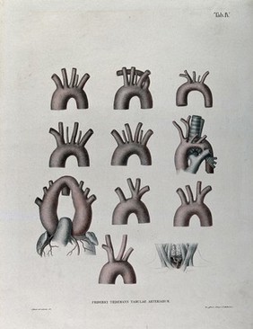
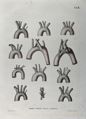
Tiedemann, Friedrich, 1781-1861 (36)
Lavater, Johann Caspar, 1741-1801 (33)
Camper, Petrus, 1722-1789 (32)
Hodges, William, 1744-1797 (32)
Schuster, Arthur, Sir, 1851-1934 (32)
Submit
4,864 results
Search result sorting
Sort by:
Relevance
Production dates
Sort order:
Ascending
Descending
Submit
Previous (page 2)
Page
3
of 163
Next (page 4)
Close modal window
Previous (page 2)
Page
3
of 163
Next (page 4)


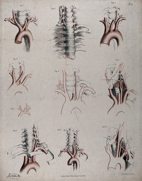


![Engravings, intended to illustrate some of the diseases of arteries. Accompanied with explanations / [Joseph Hodgson].](https://iiif.wellcomecollection.org/image/L0011706/full/600%2C/0/default.jpg)