Skip to main content
Wellcome Collection homepage
Visit us
What’s on
Stories
Collections
Get involved
About us
Sign in to your library account
Search our stories, images, catalogue and events
Library account
Search our stories, images, catalogue and events
Search
Images search
Search for images
Search
All
Stories
Images
Catalogue
Events
Colours
Licences
Creative Commons CC-BY (31)
Creative Commons CC-BY-NC (12)
In copyright (6)
Public Domain Mark (5)
Creative Commons CC0 (1)
Dates
From
to
Types/Techniques
Ephemera (4)
Collotypes (3)
Prints (3)
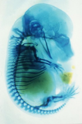
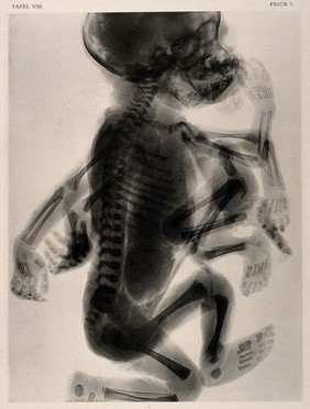
Radiographs (3)
Paintings (2)
Watercolors (2)
Engravings (1)
Subjects
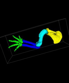
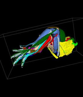
Structure (15)
Limb (12)
Support (11)
3D (9)
Aluminium (9)
Analog (9)
Analogue (9)
Artwork (9)
Black (9)
Circle (9)
Concept (9)
DEVELOPMENT (9)
Diagnose (9)
Diagnostics (9)
Digital imaging (9)
Etching (9)
Injury (9)
Knee cap (9)
Photograph (9)
Radiograph (9)
Contributors
Chiara Salvi (9)
Dr Rachel Moore (6)
NIMR, MRC (5)
A. Rueff y Cia (4)
Emei Ma, P/C Guy McLoughlin (3)
Leisewitz, Theodor, active 1908, Assistant physician of the Royal Womens Hospital, Dresden (3)
Leopold, G. (Gerhard), 1846-1911 (3)
Mike Kayser (3)
Römmler and Jonas (3)
Abigail Tucker (2)
Giles, R. H. (Robert Humphrey), 1802-1881 (2)
Kellogg Company (2)
St Bartholomew's Hospital Archives & Museum (2)
Tom Turmezei, Ken Poole and Graham Treece, University of Cambridge (2)
Buffon, Georges Louis Leclerc, comte de, 1707-1788 (1)
Chris Thorn xrayartdesign.co.uk (1)
Gautier Dagoty (1)
Godart, Thomas (1)
Paul Martin (1)
Wellcome Images (1)
Submit
55 results
Search result sorting
Sort by:
Relevance
Production dates
Sort order:
Ascending
Descending
Submit
Page
1
of 2
Next (page 2)
Close modal window
Page
1
of 2
Next (page 2)