Person
Miles, W. J. (William J.)
Images


Catalogue

- Pictures
- Online
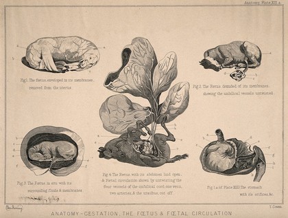
Horse foetuses: five figures showing the foetus of a horse during the gestation period, with dissections of its abdomen and stomach demonstrating the foetal circulation system. Engraving by T. Cowan after B. Herring, ca. 1860.
Herring, Benjamin, 1830-1871.Date: [1860?]Reference: 570546i- Books
Modern practical farriery : a complete system of the veterinary art as at present practised at the Royal Veterinary College, London / by W.J. Miles ; including practical treatises on cattle, their management in dairy, field, and stall / by John Walker ; pasture grasses and forage plants / by Samuel P. Preston ; the practice of sheep farming / by Charles Scott ; and the diseases of cattle, sheep, and pigs / by J.I. Lupton.
Miles, W. J. (William J.)Date: [between 1860 and 1869?]
- Books
- Online
The horse's foot, and how to keep it sound / by William Miles.
Miles, W. J. (William J.)Date: 1847
- Pictures
- Online
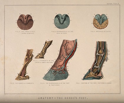
Anatomy of the horse's foot and neurotomy: six figures including the feet and hooves of a foal, an unshod horse, and the changes in form brought about by shoeing. Coloured engraving attributed to T. Cowan after a drawing attributed to B. Herring, ca. 1860.
Herring, Benjamin, 1830-1871.Date: [1860?]Reference: 570541i- Pictures
A horse's skeleton: side view, facing right. Engraving attributed to T. Cowan, after a drawing attributed to B. Herring, ca. 1860.
Herring, Benjamin, 1830-1871.Date: [1860?]Reference: 570474i