Wellcome Collection homepage
  • Visit us
  • What’s on
  • Stories
  • Collections
  • Get involved
  • About us
Sign in to your library account
Search for anything
Library account
Concept

Uterus, Pregnant

Images

  • Foetuses in utero: four figures showing cross-sections of a foetus in various positions in the uterus. Line engraving, 1791, by W. Taylor after F. Birnie, after W. Smellie.
  • Dissection of a pregnant uterus, showing the foetus at nine months, with the head positioned towards the vagina and the lower part of the placenta under the child's head, detached from the uterus. Copperplate engraving by J. Mitchel after I.V. Rymsdyk, 1774, reprinted 1851.
  • Dissection of the female abdomen, showing part of the pregnant uterus at seven months, with the surrounding viscera. Copperplate engraving by F. Aliamet after A. Cozens, 1774, reprinted 1851.
  • Three interior diagrams of an adult human uterus. Etching by G. Scotin.
  • Dissection of the pregnant female abdomen, showing the skin peeled away to reveal the swollen uterus and the viscera, side view. Copperplate engraving by G. Scotin after I.V. Rymsdyk, 1774, reprinted 1851.
All images (68)

Catalogue

    • Books
    • Online

    Anatomia uteri humani gravidi : tabulis illustrata = the anatomy of the human gravid uterus exhibited in figures / auctore Gulielmo Hunter = William Hunter.

    Hunter, William, 1718-1783. | Date: 1851
    • Pictures
    • Online

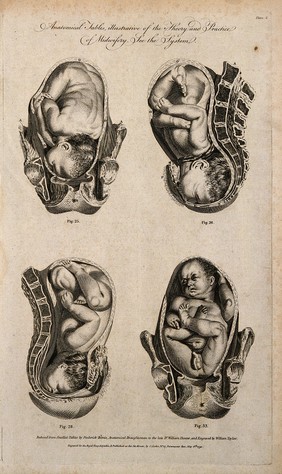
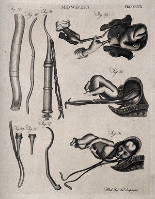
    Five diagrams illustrating the birth of a child with the use of forceps. Engraving by A. Bell.

    | Reference: 17025i
    • Pictures
    • Online

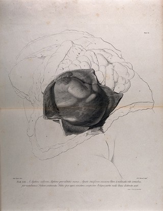
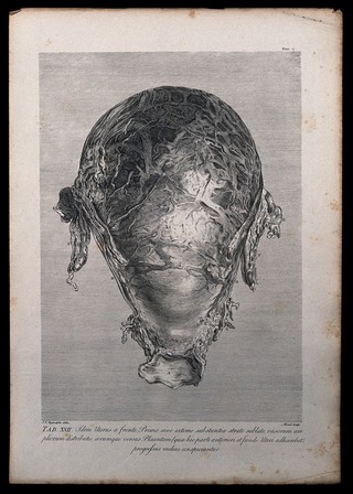
    Dissection of the pregnant uterus, showing the foetus at nine months. Copperplate engraving by R. Strange after I.V. Rymsdyk, 1774, reprinted 1851.

    Rymsdyk, Jan van, active 1750-1788. | Date: [1851] | Reference: 579806i
    Part of: Anatomia uteri humani gravidi.
    • Pictures
    • Online

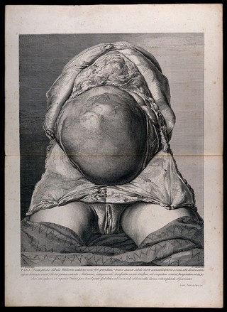
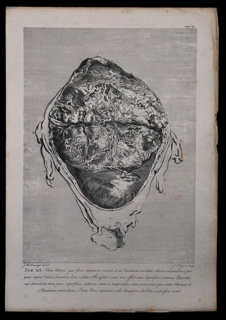
    Dissection of the pregnant uterus at five months, showing the placenta and the cervix, in relation to the bladder and urethra. Copperplate engraving by P.C. Canot after J.V. Rymsdyk, 1774, reprinted 1851.

    Rymsdyk, Jan van, active 1750-1788. | Date: [1851] | Reference: 579874i
    Part of: Anatomia uteri humani gravidi.
    • Pictures
    • Online

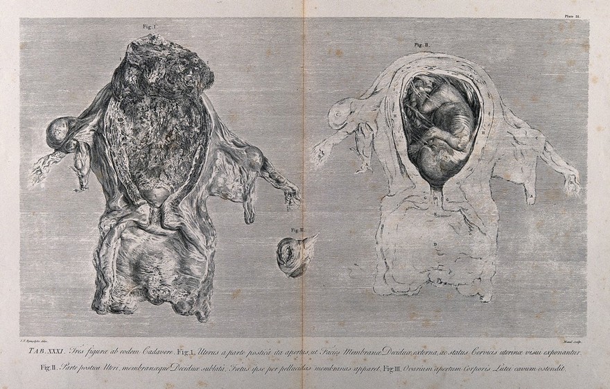
    Dissections of the pregnant uterus and the corpus luteum in the left overy, at five months: three figures. Copperplate engraving by E. Du Mesnil after J.V. Rymsdyk, 1774, reprinted 1851.

    Rymsdyk, Jan van, active 1750-1788. | Date: [1851] | Reference: 579879i
    Part of: Anatomia uteri humani gravidi.
All works (75)

Wellcome Collection

183 Euston Road
London NW1 2BE

+44 (0)20 7611 2222
info@wellcomecollection.org

  • Getting here

Today’s opening times

  • Galleries
    10:00 – 18:00
  • Library
    10:00 – 18:00
  • Café
    10:00 – 18:00
  • Shop
    10:00 – 18:00

Opening times

Our building has:

  • Step free access
  • Hearing loops

Access information

  • Visit us
  • What’s on
  • Stories
  • Collections
  • Get involved
  • About us
  • Contact us
  • Jobs
  • Media office
  • Developers
  • Privacy and terms
  • Cookie policy
  • Manage cookies
  • Modern slavery statement
Twitter
Facebook
Instagram
SoundCloud
YouTube
Tripadvisor

Except where otherwise noted, content on this site is licensed under a Creative Commons Attribution 4.0 International Licence