37 results filtered with: T cell
- Digital Images
- Online
DNA sequence of CCR5 Delta 32 gene mutation
Emei Ma, P/C Guy McLoughlin
- Digital Images
- Online
HIV assembly and budding, HIV viral life cycle, illustration
David S. Goodsell, The Scripps Research Institute
- Digital Images
- Online
Normal spleen showing B cells and T cells
Peter Lane and Fiona McConnell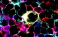
- Digital Images
- Online
Cells interacting to cause immune response
Peter Lane and Fiona McConnell
- Digital Images
- Online
Reverse transcription, HIV viral life cycle, illustration
David S. Goodsell, The Scripps Research Institute
- Digital Images
- Online
Normal spleen showing B cells and T cells
Peter Lane and Fiona McConnell
- Digital Images
- Online
HIV attachment, HIV viral life cycle, illustration
David S. Goodsell, The Scripps Research Institute
- Digital Images
- Online
Advert for Guy's Tonic, to aid indigestion and nervousness, showing a nurse pouring the tonic into a measuring cup
- Digital Images
- Online
Normal spleen showing B cells and T cells
Peter Lane and Fiona McConnell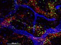
- Digital Images
- Online
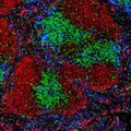
Cellular architecture of normal human skin imaged by whole mount tissue microscopy. Human skin has a rich network of white blood cells (specifically dendritic cells, T cells and macrophages) which form sheaths around blood vessels (string-like structures). A network of lymphatic vessels (ribbon-like structures) is also present. In this image, human skin lymphatic vessels (stained for LYVE-1; blue) and white blood cells comprised of dendritic cells (stained for CD11c; green) and T cells (stained for CD3; red) can be seen. Some macrophages also express the protein LYVE-1 similar to lymphatic vessel cells which can be appreciated as blue cells within and in between the sheaths of white blood cells. This normal cellular architecture is grossly disrupted in diseased skin (see related images). X10 magnification. Scale bar (white) represents 200 micrometres.
Dr. Xiao-nong Wang, Human Dendritic Cell Laboratory, Newcastle University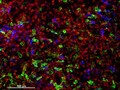
- Digital Images
- Online
Cellular architecture of human skin lymphoma imaged by whole mount tissue microscopy. Normal human skin has a rich network of white blood cells (specifically dendritic cells, T cells and macrophages) which form sheaths around blood vessels. In diseased skin, such as in skin lymphoma as seen here, this normal architecture becomes distorted. In this image, lots of T cells (stained for CD3; red), dendritic cells (stained for CD11c; green) and macrophages (stained for LYVE-1; blue) have infiltrated the skin. X20 magnification. Scale bar (white) represents 100 micrometres.
Dr. Xiao-nong Wang, Human Dendritic Cell Laboratory, Newcastle University
- Digital Images
- Online
Normal spleen showing B cells and T cells
Peter Lane and Fiona McConnell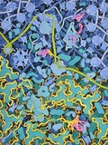
- Digital Images
- Online
HIV transcription, HIV viral life cycle, illustration
David S. Goodsell, The Scripps Research Institute
- Digital Images
- Online
HIV and antibodies, HIV viral life cycle, illustration
David S. Goodsell, The Scripps Research Institute
- Digital Images
- Online
NK T-cell lymphoma is a highly aggressive cancer of a specific type of immune cell called lymphoid cells, and is associated with the Epstein Barr virus (glandular fever). In later stages of the disease, the lymphoma can spread to the lymph nodes, as in this case.
William R. Geddie
- Digital Images
- Online
Cellular architecture of normal human skin imaged by whole mount tissue microscopy. Human skin has a rich network of white blood cells (specifically dendritic cells, T cells and macrophages) which form sheaths around blood vessels. This image was taken less than 20 micrometres beneath the junction that joins the dermal and epidermal layers of the skin (dermo-epidermal junction). At this level, dendritic cells (stained for CD11c; green) form clusters around and between blood capillary loops (stained for CD31; red). The blind-ended tips of initial lymphatic vessels are just visible (stained for LYVE-1; blue) at this level. This normal cellular architecture is grossly disrupted in diseased skin (see related images). Scale bar (white) represents 200 micrometres.
Dr. Xiao-nong Wang, Human Dendritic Cell Laboratory, Newcastle University
- Digital Images
- Online
Cellular architecture of normal human skin imaged by whole mount tissue microscopy. Human skin has a rich network of white blood cells (specifically dendritic cells, T cells and macrophages) which form sheaths around blood vessels. In this image, T cells (stained for CD3; red) dendritic cells (stained for MHC class II; green) and macrophages (stained for LYVE-1; blue with some cells showing a tinge of green) can be seen. Cell nuclei have been stained with DAPI (grey). This normal cellular architecture is grossly disrupted in diseased skin (see related images). X10 magnification. Scale bar (white) represents 200 micrometres.
Dr. Xiao-nong Wang, Human Dendritic Cell Laboratory, Newcastle University
- Digital Images
- Online
DNA sequence of CCR5 Delta 32 gene mutation
Emei Ma, P/C Guy McLoughlin
- Digital Images
- Online
St George and the dragon - T lymphocyte killing a cancer cell
Odra Noel
- Digital Images
- Online
Cellular architecture of normal human skin imaged by whole mount tissue microscopy. Human skin has a rich network of white blood cells (specifically dendritic cells, T cells and macrophages) which form sheaths around blood vessels. In this image, T cells (stained for CD3; red) dendritic cells (stained for MHC class II; green) and macrophages (stained for LYVE-1; blue with some cells showing a tinge of green) can be seen. Cell nuclei have been stained with DAPI (grey). This normal cellular architecture is grossly disrupted in diseased skin (see related images). X20 magnification. Scale bar (white) represents 100 micrometres.
Dr. Xiao-nong Wang, Human Dendritic Cell Laboratory, Newcastle University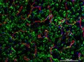
- Digital Images
- Online
Cellular architecture of normal human skin imaged by whole mount tissue microscopy. Human skin has a rich network of white blood cells (specifically dendritic cells, T cells and macrophages) which form sheaths around blood vessels. This image was taken directly beneath the junction that joins the dermal and epidermal layers of the skin (dermo-epidermal junction). At this level, the capillary network (stained for CD31; red) is visualised against a lawn of autofluorescent dermal papillae (finger-like projections of the dermis; green) scattered with dendritic cells (stained for CD11c; green) and macrophages (stained for LYVE-1; blue). This normal cellular architecture is grossly disrupted in diseased skin (see related images). Scale bar (white) represents 200 micrometres.
Dr. Xiao-nong Wang, Human Dendritic Cell Laboratory, Newcastle University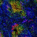
- Digital Images
- Online
Normal spleen showing B cells and T cells
Peter Lane and Fiona McConnell
- Digital Images
- Online
Cellular architecture of normal human skin imaged by whole mount tissue microscopy. Human skin has a rich network of white blood cells (specifically dendritic cells, T cells and macrophages) which form sheaths around blood vessels. In this image, blood vessels (string-like structures stained for CD31; green), lymphatic vessels (ribbon-like structures stained for LYVE-1; blue) and T cells (stained for CD3; red) can be seen. T cells are only found around dermal blood vessels. Macrophages (stained for LYVE-1; blue) are also present. This normal cellular architecture is grossly disrupted in diseased skin (see related images). X10 magnification. Scale bar (white) represents 200 micrometres.
Dr. Xiao-nong Wang, Human Dendritic Cell Laboratory, Newcastle University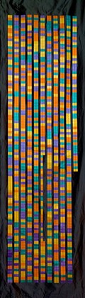
- Digital Images
- Online
DNA sequence of CCR5 Delta 32 gene mutation
Emei Ma, P/C Guy McLoughlin
- Digital Images
- Online
HIV maturation, HIV viral life cycle, illustration
David S. Goodsell, The Scripps Research Institute