19 results filtered with: Double helix

- Digital Images
- Online
Shadow of a DNA double helix on sequencing output
Peter Artymiuk
- Digital Images
- Online
DNA double helix
Peter Artymiuk
- Digital Images
- Online
Illustration of the DNA double helix. The sugar-phosphate backbone of the two complementary strands are visible (red and blue).
Susan Lockhart
- Digital Images
- Online
Model of a DNA double helix according to the correct dimensions of the natural molecule.
Peter Artymiuk
- Digital Images
- Online
Holliday junction and proteins
John Rafferty
- Digital Images
- Online
Model of a DNA double helix according to the correct dimensions of the natural molecule.
Peter Artymiuk
- Digital Images
- Online
53 in the form of a double helix
Peter Artymiuk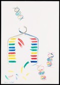
- Digital Images
- Online
Illustration depicting semi-conservative DNA replication. A DNA double helix prior to replication is shown in the top left of the image. The sugar phosphate backbone and nucleotide bases are visible. Complementary base pairing of adenine with thymine (blue with green) and guanine with cytosine (red with yellow) is shown. During replication, a length of the double helix temporarily unwinds and separates into two strands. Free nucleotides bind by complementary base pairing to the recently exposed nucleotides on each strand which act as a template. Two new double helices are formed, each containing one original generation and one new generation strand of DNA. The sequence of base pairs in each double helix is identical to the original.
Susan Lockhart
- Digital Images
- Online
DNA double helix and sequencing output
Peter Artymiuk
- Digital Images
- Online
DNA double helix and sequencing output
Peter Artymiuk
- Digital Images
- Online
DNA double helix and sequencing output
Peter Artymiuk
- Digital Images
- Online
Illustration depicting semi-conservative DNA replication. Three generations of DNA are shown. After separation of the DNA double helix, two new complementary DNA strands are synthesised (indicated by a new colour). Complementary base pairing and hydrogen bonding results in formation of a new double helix.
Susan Lockhart
- Digital Images
- Online
Spirals of DNA molecules
Annie Cavanagh
- Digital Images
- Online
DNA double helix and sequencing output
Peter Artymiuk
- Digital Images
- Online
DNA double helix
Peter Artymiuk
- Digital Images
- Online
Model of a DNA double helix according to the correct dimensions of the natural molecule.
Peter Artymiuk
- Digital Images
- Online
RuvA protein bound to DNA - side view
John Rafferty
- Digital Images
- Online
DNA double helix and sequencing output
Peter Artymiuk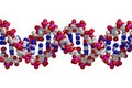
- Digital Images
- Online
DNA double helix
Peter Artymiuk