30 results filtered with: Brain - Localisation of functions
- Pictures
The brain seen from the underside, sectioned horizontally; with attention to the parts associated by Hollander's system of phrenology with the faculties of external perception and its memory. Process print, 1901, after etching, 1809.
Date: 1901Reference: 28018i- Pictures
Two skulls with temporal regions of different size and shape. Photomechanical reproduction with painting, c. 1902.
Date: 1902Reference: 28067i
- Pictures
- Online
Diagram of the brain for a phrenological textbook. Pen drawing, c. 1902.
Date: 1902Reference: 27968i
- Pictures
- Online
A male figure and three phrenological heads. Wood engraving.
Reference: 27704i
- Pictures
- Online
The human brain, divided according to Bernard Hollander's system of phrenology. Process print with pen and ink, c. 1902.
Date: [approximately 1902]Reference: 27959i
- Pictures
- Online
A male brain, sectioned vertically. Process print, 1901, after etching, 1809.
Date: 1901Reference: 28021i
- Pictures
- Online
Right profile of head with depressed frontal lobes, divided up to show the location of all the lobes. Drawing, c. 1900.
Date: c. 1900Reference: 28386i
- Pictures
- Online
A female brain, sectioned vertically: side view. Process print, 1901, after etching, 1809.
Date: 1901Reference: 28022i
- Pictures
- Online
Phrenological chart; with design of head containing symbols of the phrenological 'faculties'. Etching after O.S. Fowler (?).
Fowler, O. S. (Orson Squire), 1809-1887.Reference: 28451i
- Pictures
- Online
Skull of a soldier: frontal view. Lithograph, 1835.
Date: 1835Reference: 28075i
- Pictures
- Online
Left profile of a head showing depressed frontal lobes. Drawing, c. 1900.
Date: c. 1900Reference: 28387i- Pictures
The brain: side view illustrating the distance of the occipital bone from the phrenological 'organ of philoprogenitiveness'. Process print, 1901, after etching, 1809.
Date: 1901Reference: 28019i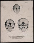
- Pictures
- Online
The bases of three skulls: a new born infant's, a misogynist's, and a man suffering from satyriasis. Process print, 1901, after etching, 1809.
Date: 1901Reference: 28023i- Pictures
The brain, sectioned vertically; showing the sites of some phrenological faculties. Process print, 1901, after etching, 1809.
Date: 1901Reference: 28020i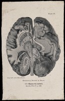
- Pictures
- Online
The brain seen from the underside, sectioned horizontally; with attention to the part associated by Hollander's system of phrenology with memory for numbers. Process print, 1901, after etching, 1809.
Date: 1901Reference: 28017i- Pictures
A brain seen from the underside; with attention to the area associated by Gall with verbal memory. Process print, 1901, after etching, 1809.
Date: 1901Reference: 28011i
- Pictures
- Online
Phrenological chart with three figures of a head and sketches of the heads of famous men. Coloured lithograph, 1836.
Date: 1836Reference: 28571i
- Pictures
- Online
Child's head with large temporal lobes and depressed frontal lobe. Drawing, c. 1900.
Date: c. 1900Reference: 28388i
- Pictures
- Online
Two sections of the brain, divided into different lobes and faculties, according to Hollander's system of phrenology. Pen drawing, c. 1902.
Date: 1902Reference: 27965i
- Pictures
- Online
The human head, divided according to the system of phrenology. Coloured lithograph by C. Ingrey, 1824.
Date: 1824Reference: 27671i
- Pictures
- Online
Head of woman showing musical ability, according to phrenological classification. Drawing, c. 1900.
Date: c. 1900Reference: 28389i
- Pictures
- Online
A head divided into thirty seven compartments, each containing an image representing a phrenological faculty. Wood engraving, after O.S. Fowler, c. 1840.
Fowler, O. S. (Orson Squire), 1809-1887.Reference: 27714i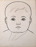
- Pictures
- Online
Head of a child with large cheeks. Drawing, c. 1900.
Date: c. 1900Reference: 28390i
- Pictures
- Online
Three heads showing phrenological traits associated with insanity: a mentally defective person, a mad woman, and the murderer P.F. Lacenaire. Lithograph by C. Picard, 1842, after J.P. Thenot.
Thénot, J. P. (Jean Pierre), 1803-1857.Date: 1842Reference: 27673i
- Pictures
- Online
Head showing the 'convolutions' (lobes) of the brain. Pen drawing after R. W. Reid.
Reference: 28366i