123 results
- Archives and manuscripts
`Illustrations and graphs of my calculations for capillary surface area (not published)'
Date: Mid 20th centuryReference: PP/DBD/B/30Part of: Daly, Professor Ivan de Burgh- Books
Patna City municipality, surface drainage construction : area blocks 36 and 37 : report / [F. C. Temple].
Temple, F. C. (Frederick Charles)Date: [1919]- Books
Possible relations of the weight of the lungs and other organs to body-weight and surface area (in dogs) / by G.N. Stewart.
Stewart, G. N. (George Neil), 1860-1930.Date: [1921?]
- Digital Images
- Online
Inner surface of ileum
Prof Giorgio Gabella
- Digital Images
- Online
Leucoma on the tongue with fluffy surface
Godart, Thomas
- Books
- Online
The blood volume of mammals as determined by experiments upon rabbits, guinea-pigs, and mice : and its relationship to the body weight and to the surface area expressed in a formula / by Georges Dreyer and William Ray.
Dreyer, G. (Georges), 1873-1934.Date: [1910]
- Pictures
- Online
Philadelphia College of Pharmacy and Science: pharmacy area for students. Photograph, c. 1933.
Date: c. 1933Reference: 30236i
- Digital Images
- Online
Cone rich area of the retina - 3D image
Chris Guerin
- Digital Images
- Online
Intestinal villi
Liz Hirst, Medical Research Council
- Digital Images
- Online
Villi in the small intestine
Paul Appleton, University of Dundee
- Digital Images
- Online
Human small intestine showing villi. The cytokeratinin the cells is stained blue, the cell nuclei are stained red and the endothelial cells lining the blood vessels are stained green.
S. Schuller
- Digital Images
- Online
Villi in the small intestine
Paul Appleton, University of Dundee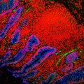
- Digital Images
- Online
Human small intestine showing villi and glands. The cytokeratinin the cells is stained blue, the cell nuclei are stained red and the endothelial cells lining the blood vessels are stained green.
S. Schuller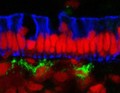
- Digital Images
- Online
Human small intestine showing the columnar epithelium. The cytokeratinin the cells is stained blue, the cell nuclei are stained red and the endothelial cells lining the blood vessels are stained green.
S. Schuller
- Digital Images
- Online
Human small intestine showing villi and glands. The cytokeratinin the cells is stained blue, the cell nuclei are stained red and the endothelial cells lining the blood vessels are stained green.
S. Schuller
- Digital Images
- Online
Human small intestine showing villi and glands. The cytokeratin in the cells is stained blue, the cell nuclei are stained red and the endothelial cells lining the blood vessels are stained green.
S. Schuller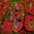
- Digital Images
- Online
Mouse colon infected with Citrobacter rodentium
S. Schuller
- Digital Images
- Online
Mouse colon infected with Citrobacter rodentium
S. Schuller
- Digital Images
- Online
Neuroendocrine cells in the small intestine
S. Schuller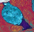
- Digital Images
- Online
Goblet cells are packed full of mucous globules (blue), which they release to provide lubrication and protection to the inner surfaces of the intestine and the respiratory system among others. The mucous globules are condensed inside the goblet cell but expand hugely once they are released, absorbing water within 20 milliseconds. This rapid release occurs in response to lots of different stimuli and allows the mucous to get to work instantly.
University of Edinburgh
- Digital Images
- Online
A single enteropathogenic E. coli in the intestine
S. Schuller
- Digital Images
- Online
Surface of the corneal epithelium - coloured
Rob Young- Archives and manuscripts
- Online
Dissertation in Draft: Appendix II
Date: 1953Reference: PP/CRI/F/1/16Part of: Francis Crick (1916-2004): archives
- Digital Images
- Online
Gut
Kevin Mackenzie, University of Aberdeen
- Digital Images
- Online
Villus from the small intestine
S. Schuller