52 results
- Archives and manuscripts
Acrocyanosis
Date: 1894-1954Reference: PP/FPW/B.1Part of: Parkes Weber, Frederick (1863-1962)- Books
The immunological basis of connective tissue disorders : proceedings of the fifth Lepetit Colloquium, held in Madrid, Spain, 11-13 November 1974 / editor Luigi G. Silvestri.
Lepetit Colloquium 1974 : Madrid, Spain)Date: 1975
- Books
- Online
Three cases of tumour arising from skin-glands in the dog : showing the connection between disorder of the glandular structure and function and cancerous invasion of the connective tissue / by Charles Creighton.
Creighton, Charles, 1847-1927.Date: 1882- Archives and manuscripts
Visits and conferences 'Chronic Rheumatism'
Date: 1926-1928Reference: SA/HEB/D/2/1Part of: Heberden Collection- Books
Practical genetic counselling / Peter S. Harper.
Harper, Peter S.Date: 1993- Archives and manuscripts
- Online
Offprints: Respiration
Date: 1959-1967Reference: PP/CAT/C.1Part of: Dr Mary Catterall
- Digital Images
- Online
Cellular architecture of normal human skin imaged by whole mount tissue microscopy. Human skin has a rich network of white blood cells (specifically dendritic cells, T cells and macrophages) which form sheaths around blood vessels. In this image, T cells (stained for CD3; red) dendritic cells (stained for MHC class II; green) and macrophages (stained for LYVE-1; blue with some cells showing a tinge of green) can be seen. Cell nuclei have been stained with DAPI (grey). This normal cellular architecture is grossly disrupted in diseased skin (see related images). X10 magnification. Scale bar (white) represents 200 micrometres.
Dr. Xiao-nong Wang, Human Dendritic Cell Laboratory, Newcastle University
- Digital Images
- Online
Cellular architecture of normal human skin imaged by whole mount tissue microscopy. Human skin has a rich network of white blood cells (specifically dendritic cells, T cells and macrophages) which form sheaths around blood vessels. In this image, T cells (stained for CD3; red) dendritic cells (stained for MHC class II; green) and macrophages (stained for LYVE-1; blue with some cells showing a tinge of green) can be seen. Cell nuclei have been stained with DAPI (grey). This normal cellular architecture is grossly disrupted in diseased skin (see related images). X20 magnification. Scale bar (white) represents 100 micrometres.
Dr. Xiao-nong Wang, Human Dendritic Cell Laboratory, Newcastle University
- Digital Images
- Online
Cellular architecture of normal human skin imaged by whole mount tissue microscopy. Human skin has a rich network of white blood cells (specifically dendritic cells, T cells and macrophages) which form sheaths around blood vessels. In this image, blood vessels (string-like structures stained for CD31; red), lymphatic vessels (ribbon-like structures stained for LYVE-1; blue) and dendritic cells (stained for CD11c; green) can be seen. Macrophages (stained for LYVE-1; blue) are also present. This normal cellular architecture is grossly disrupted in diseased skin (see related images). X10 magnification. Scale bar (white) represents 200 micrometres.
Dr. Xiao-nong Wang, Human Dendritic Cell Laboratory, Newcastle University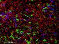
- Digital Images
- Online
Cellular architecture of human skin lymphoma imaged by whole mount tissue microscopy. Normal human skin has a rich network of white blood cells (specifically dendritic cells, T cells and macrophages) which form sheaths around blood vessels. In diseased skin, such as in skin lymphoma as seen here, this normal architecture becomes distorted. In this image, lots of T cells (stained for CD3; red), dendritic cells (stained for CD11c; green) and macrophages (stained for LYVE-1; blue) have infiltrated the skin. X20 magnification. Scale bar (white) represents 100 micrometres.
Dr. Xiao-nong Wang, Human Dendritic Cell Laboratory, Newcastle University
- Digital Images
- Online
Cellular architecture of normal human skin imaged by whole mount tissue microscopy. Human skin has a rich network of white blood cells (specifically dendritic cells, T cells and macrophages) which form sheaths around blood vessels. In this image, blood vessels (string-like structures stained for CD31; green), lymphatic vessels (ribbon-like structures stained for LYVE-1; blue) and T cells (stained for CD3; red) can be seen. T cells are only found around dermal blood vessels. Macrophages (stained for LYVE-1; blue) are also present. This normal cellular architecture is grossly disrupted in diseased skin (see related images). X10 magnification. Scale bar (white) represents 200 micrometres.
Dr. Xiao-nong Wang, Human Dendritic Cell Laboratory, Newcastle University
- Digital Images
- Online
Cellular architecture of normal human skin imaged by whole mount tissue microscopy. Human skin has a rich network of white blood cells (specifically dendritic cells, T cells and macrophages) which form sheaths around blood vessels. This image was taken greater than 150 micrometres beneath the junction that joins the dermal and epidermal layers of the skin (dermo-epidermal junction). At this level, dendritic cells (stained for CD11c; green) and macrophages (stained for LYVE-1; blue) form clusters around blood vessels (stained for CD31; red). This normal cellular architecture is grossly disrupted in diseased skin (see related images). Scale bar (white) represents 100 micrometres.
Dr. Xiao-nong Wang, Human Dendritic Cell Laboratory, Newcastle University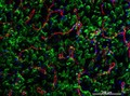
- Digital Images
- Online
Cellular architecture of normal human skin imaged by whole mount tissue microscopy. Human skin has a rich network of white blood cells (specifically dendritic cells, T cells and macrophages) which form sheaths around blood vessels. This image was taken directly beneath the junction that joins the dermal and epidermal layers of the skin (dermo-epidermal junction). At this level, the capillary network (stained for CD31; red) is visualised against a lawn of autofluorescent dermal papillae (finger-like projections of the dermis; green) scattered with dendritic cells (stained for CD11c; green) and macrophages (stained for LYVE-1; blue). This normal cellular architecture is grossly disrupted in diseased skin (see related images). Scale bar (white) represents 200 micrometres.
Dr. Xiao-nong Wang, Human Dendritic Cell Laboratory, Newcastle University
- Digital Images
- Online
Cellular architecture of normal human skin imaged by whole mount tissue microscopy. Human skin has a rich network of white blood cells (specifically dendritic cells, T cells and macrophages) which form sheaths around blood vessels. This image was taken less than 20 micrometres beneath the junction that joins the dermal and epidermal layers of the skin (dermo-epidermal junction). At this level, dendritic cells (stained for CD11c; green) form clusters around and between blood capillary loops (stained for CD31; red). The blind-ended tips of initial lymphatic vessels are just visible (stained for LYVE-1; blue) at this level. This normal cellular architecture is grossly disrupted in diseased skin (see related images). Scale bar (white) represents 200 micrometres.
Dr. Xiao-nong Wang, Human Dendritic Cell Laboratory, Newcastle University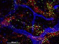
- Digital Images
- Online
Cellular architecture of normal human skin imaged by whole mount tissue microscopy. Human skin has a rich network of white blood cells (specifically dendritic cells, T cells and macrophages) which form sheaths around blood vessels (string-like structures). A network of lymphatic vessels (ribbon-like structures) is also present. In this image, human skin lymphatic vessels (stained for LYVE-1; blue) and white blood cells comprised of dendritic cells (stained for CD11c; green) and T cells (stained for CD3; red) can be seen. Some macrophages also express the protein LYVE-1 similar to lymphatic vessel cells which can be appreciated as blue cells within and in between the sheaths of white blood cells. This normal cellular architecture is grossly disrupted in diseased skin (see related images). X10 magnification. Scale bar (white) represents 200 micrometres.
Dr. Xiao-nong Wang, Human Dendritic Cell Laboratory, Newcastle University
- Pictures
- Online
Female genitalia showing severely diseased tissue of the labia, and some sores on the thighs. Watercolour by Christopher D' Alton.
D'Alton, Christopher, active 1847-1871.Date: 1800-1899Reference: 38308i- Archives and manuscripts
Scientific Basis of Medicine Series 7 (C.342)
Date: 1976-1977Reference: GC/170/13/1Part of: University Of London Audio-visual Centre
- Digital Images
- Online
Section of lung exhibiting malignant disease, probably lympho-sarcoma
Godart, Thomas- Archives and manuscripts
Heberden Society
Date: 1714-1983Reference: SA/HEB/APart of: Heberden Collection- Books
Miscellaneous cases and commentaries / by Jonathan Hutchinson ..., Surgeon to the London Hospital and Lecturer on Surgery; Surgeon to the London Hospital for Diseases of the Skin, and Assistant Surgeon to the Royal Ophthalmic Hospital.
Hutchinson, Jonathan, Sir, 1828-1913Date: 1868- Archives and manuscripts
Abstracts of academic articles submitted to the Heberden society
Date: Nov 1982Reference: SA/HEB/A/1/9Part of: Heberden Collection
- Digital Images
- Online
Acute inflammation of the larynx
Delamotte, William Alfred- Archives and manuscripts
Fell, Dame Honor Bridget (1900-1986)
Fell, Dame Honor Bridget, 1900-1986Date: 1919-1988Reference: PP/HBF- Pictures
Myositis ossificans in hemiplegia, in a 74-year old woman: detail sketch of swelling below right groin. Watercolour by Barbara E. Nicholson, 1953.
Nicholson, BarbaraDate: 1953Reference: 34744iPart of: Barbara Nicholson medical illustration collection.- Books
The custom-made brain : cerebral plasticity, regeneration, and enhancement / Jean-Didier Vincent and Pierre-Marie Lledo ; translated by Laurence Garey.
Vincent, Jean-DidierDate: [2014]