31 results
- Digital Images
- Online
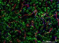
Cellular architecture of normal human skin imaged by whole mount tissue microscopy. Human skin has a rich network of white blood cells (specifically dendritic cells, T cells and macrophages) which form sheaths around blood vessels. This image was taken directly beneath the junction that joins the dermal and epidermal layers of the skin (dermo-epidermal junction). At this level, the capillary network (stained for CD31; red) is visualised against a lawn of autofluorescent dermal papillae (finger-like projections of the dermis; green) scattered with dendritic cells (stained for CD11c; green) and macrophages (stained for LYVE-1; blue). This normal cellular architecture is grossly disrupted in diseased skin (see related images). Scale bar (white) represents 200 micrometres.
Dr. Xiao-nong Wang, Human Dendritic Cell Laboratory, Newcastle University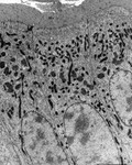
- Digital Images
- Online
Gut
Kevin Mackenzie, University of Aberdeen
- Digital Images
- Online
Osteocytes
Kevin Mackenzie, University of Aberdeen
- Digital Images
- Online
Neuropil
Prof. Bill Harris
- Digital Images
- Online
Human neural stem cells stained for Sox2 (green) and vimentin (red). Both are markers of neural stem cells.
Yirui Sun
- Digital Images
- Online
Mouse neural stem cells growing in culture. Neural stem cells can be made to develop into cells found in the central nervous system; neurons, astrocytes and oligodendrocytes.
Yirui Sun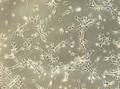
- Digital Images
- Online
Human neural stem cells growing in culture. Neural stem cells can be made to develop into cells found in the central nervous system; neurons, astrocytes and oligodendrocytes.
Yirui Sun
- Digital Images
- Online
Skin cancer cell
Annie Cavanagh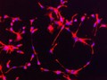
- Digital Images
- Online
Human neural stem cells stained for nestin (red). Nestin is a type of intermediate filamant protein that is used as a marker of neural stem cells. The blue dots are the cell nuclei stained with DAPI. Neural stem cells can be made to develop into cells found in the central nervous system; neurons, astrocytes and oligodendrocytes.
Yirui Sun
- Digital Images
- Online
Mouse neural stem cells growing in culture. Neural stem cells can be made to develop into cells found in the central nervous system; neurons, astrocytes and oligodendrocytes.
Yirui Sun
- Digital Images
- Online
Embryonic kidney cells
Annie Cavanagh
- Digital Images
- Online
Embryonic kidney cells
Annie Cavanagh- Books
Molecular, cellular, biological characterization of childhood thyroid cancer : final report / editors E.D. Williams and N.D. Tronko.
Date: 1996
- Digital Images
- Online
Macrophage/lymphocyte/fibroblast interaction
Rob Young- Archives and manuscripts
Alan Frederick Williams (1945-1992): archive
Williams, Alan Frederick, PhD, FRS, (1945-1992), biochemist and immunologist, Director Medical Research Council Cellular Immunology Unit 1977-1992Date: c1960s-1992Reference: PP/AFW
- Digital Images
- Online
Cervical Cancer
Kate Cragoe Mayfield
- Digital Images
- Online
Human kidney cell, Gated-STED microscopy
Alison Dun, ESRIC (Edinburgh Super-Resolution Imaging Consortium)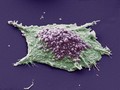
- Digital Images
- Online
Lung cancer cell. This image shows a single cell grown from a culture of lung epithelial carcinoma (cancer) cells. The purple area shows blebbing.
Anne Weston, Francis Crick Institute
- Digital Images
- Online
Prostate cancer cell spheroid, SEM
Izzat Suffian, David McCarthy & Khuloud T. Al-Jamal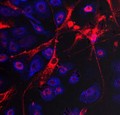
- Digital Images
- Online
Stem cell transfer of mitochondria
Queen's University Belfast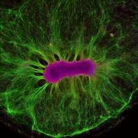
- Digital Images
- Online
Brain Organoid.
Edington, Collin.Date: 2017
- Digital Images
- Online
Situs inversus, illustration
S. Roy
- Digital Images
- Online
Villi in the small intestine
Paul Appleton, University of Dundee
- Digital Images
- Online
The Ebola virus
Odra Noel
- Digital Images
- Online
The Ebola virus
Odra Noel